. 2022 May;381(2):120-128.
doi: 10.1124/jpet.121.001034. Epub 2022 Mar 19.

High Salt Upregulates Ca2+-Sensing Receptor Expression and Ca2+-Induced Relaxation of Contracted Mesenteric Arteries from Dahl Salt-Sensitive Rats

Affiliations

High Salt Upregulates Ca2+-Sensing Receptor Expression and Ca2+-Induced Relaxation of Contracted Mesenteric Arteries from Dahl Salt-Sensitive Rats

Lakeesha E Bridges et al. J Pharmacol Exp Ther. 2022 May.

Abstract

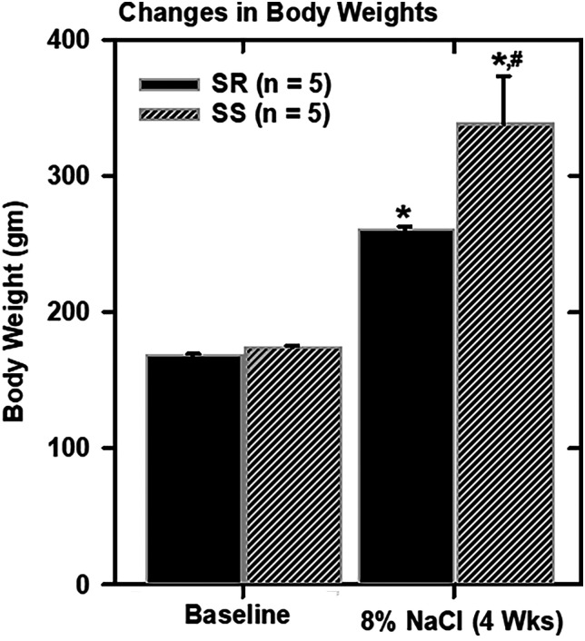
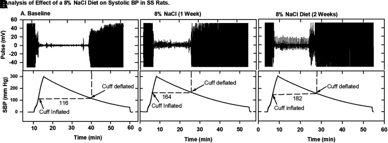
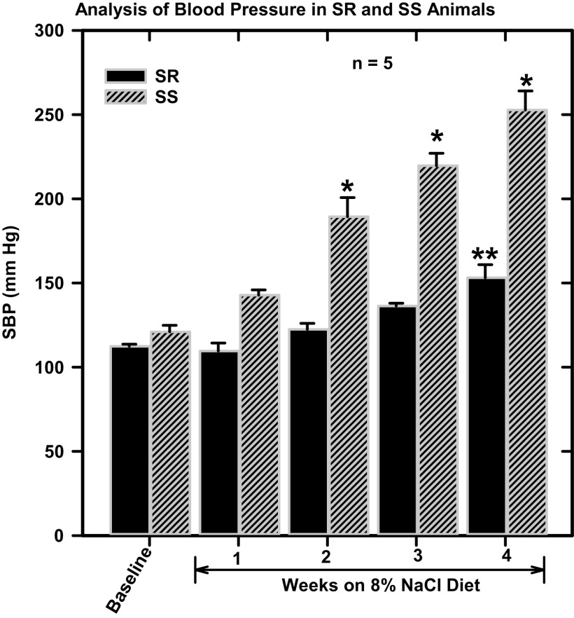
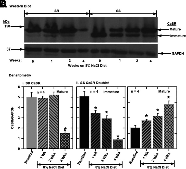
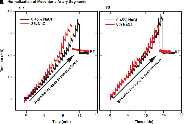
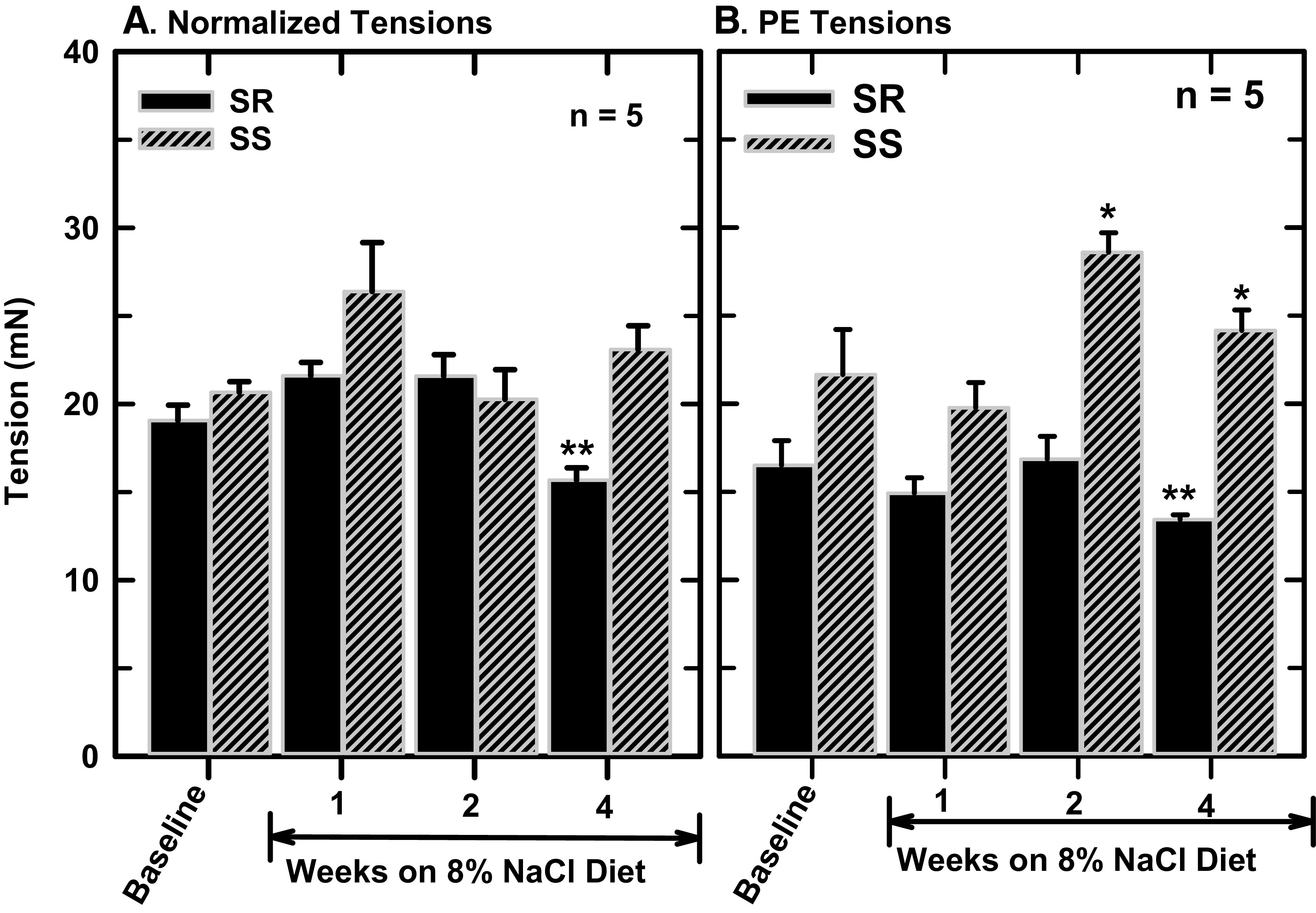
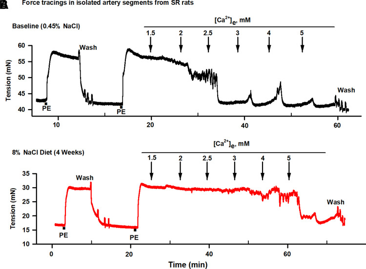
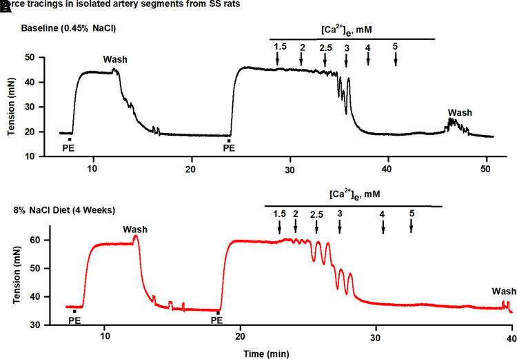
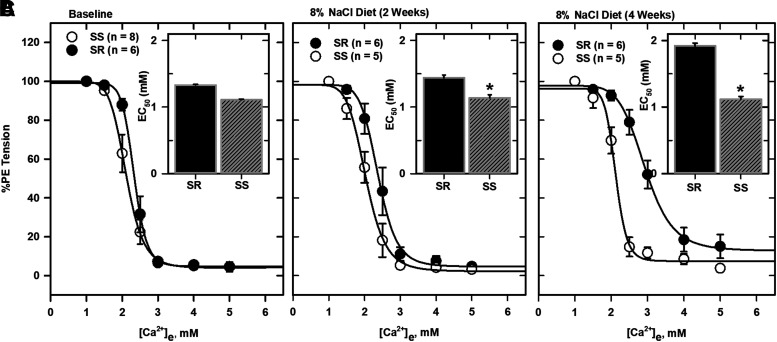
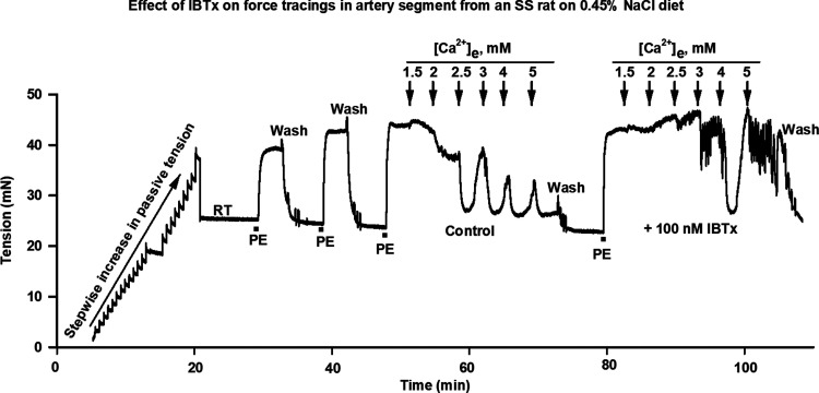
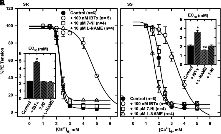
High Ca2+ lowers blood pressure in hypertension, but the mechanism is not clear. The missing link may be the perivascular sensory nerve Ca2+-sensing receptor (CaSR) that mediates a vasodilator system after activation by interstitial Ca2+ Our results show that high salt increased CaSR expression in mesenteric arteries as well as Ca2+ relaxation of contracted mesenteric arteries from salt-sensitive (SS) rats. The CaSR was expressed as a doublet (≈120-150 kDa) in arteries from animals fed a high-salt diet for 1-4 weeks. The higher molecular weight glycosylated protein increased in arteries from SS animals; however, expression of the low molecular mass high-mannose protein decreased over 4 weeks of feeding the diet. In tissues from salt-resistant (SR) rats, the diet decreased CaSR expression after 4 weeks. Ca2+ relaxation of mesenteric arteries under phenylephrine tone increased in SS rats but decreased in arteries from SR rats fed the high-salt diet. Ca2+-activated K+ channels have a larger role in Ca2+ relaxation of arteries in SR than SS rats. The data suggest that high salt epigenetically regulates the receptor at the translational level in vivo and that the in vitro effect of Ca2+ is on receptor trafficking and signaling. In conclusion, upregulated expression of the CaSR in salt sensitivity increased receptor-mediated vascular relaxation. These findings show that CaSR signaling may compensate for changes in the vasculature in salt-sensitive hypertension. SIGNIFICANCE STATEMENT: The perivascular sensory nerve Ca2+-sensing receptor (CaSR) mediates Ca2+ relaxation of isolated mesenteric arteries under tension. This receptor may therefore play a significant role in relaxation of resistance arteries in vivo, thus explaining the blood pressure-lowering effect of dietary Ca2+. The present studies describe the effect of high salt-induced upregulation of the CaSR in salt-sensitive rats and the roles played by Ca2+-activated K+ channels and nitric oxide in Ca2+ responses.


Figures

Fig. 1.
Fig. 1.
Effect of an 8% NaCl diet on body weights of SR and SS rats. Body weights of animals fed the high-salt diet for 4 weeks. Data plotted are means (± S.E.M.) of determinations in five animals as shown. *Significantly different from baseline values; #Significantly different from SR.
Fig. 2.
Fig. 2.
Effect of an 8% NaCl diet on BP in SS rats. Animals were fed the high-salt diet for 1–2 weeks, and SBPs were determined by a computerized tail-cuff method. (A) Baseline pulse and SBP recordings in an SS rat before initiation of diet treatment and after feeding of the 8% NaCl diet for (B) 1 week or (C) 2 weeks. Cuff inflations and deflations are shown on the tracings.
Fig. 3.
Fig. 3.
Analysis of the effects of an 8% NaCl diet on BP in SR and SS rats. (A) Baseline pulse and SBPs, (B) 8% NaCl diet for 1 week, and (C) 8% NaCl diet for 2 weeks in SS animals. SBPs were analyzed after feeding of the high-salt diet for 1–4 weeks to SR and SS animals. Data are means (± S.E.M.) of determinations in five animals from each group. *Significantly different from baseline and SR; **Significantly different from SR baseline.
Fig. 4.
Fig. 4.
Effect of an 8% NaCl diet on expression of the CaSR in mesenteric arteries from SR and SS rats. Animals were fed the high-salt diet for over 4 weeks, and protein extracts from mesenteric arteries were analyzed by Western blotting with a polyclonal CaSR antibody (PA1-37213) and compared with baseline expressions. (A) Western blot analysis of protein extracts from mesenteric arteries from SR and SS rats. (B) Bar charts showing densitometry analysis of the high molecular mass CaSR band normalized to polyclonal GAPDH antibody (FL-335) as the loading standard; (i) analysis of protein extracts of mesenteric arteries from SR rats showing expression of the high molecular mass, glycosylated, mature CaSR; (ii) analysis of protein extracts of mesenteric arteries from SS rats showing expression of the high molecular, glycosylated, mature CaSR and the low molecular mass, high-mannose, immature CaSR. Data are means (± S.E.M.) of four animals. *Significantly different from baseline.
Fig. 5.
Fig. 5.
Effect of an 8% NaCl diet on “normalization” of mounted mesenteric arteries from SR and SS rats. Segments (2 mm) of isolated mesenteric arteries were mounted in a wire myograph chamber in PSS medium (with 1 mM Ca2+ and 100 μM ascorbic acid), equilibrated at 37°C with aeration (95% air/5% CO2), and “normalized” by stepwise increases in passive force (in the absence of smooth muscle activation) until a resting tension (RT) is achieved. “Normalization” of mounted vessels from (A) SR rats at baseline and (B) SS rats at baseline (on a 0.45% NaCl diet) or on 8% NaCl diet for 4 weeks.
Fig. 6.
Fig. 6.
Effect of an 8% NaCl diet on normalized and PE tensions in mesenteric arteries. Normalized tensions and tensions generated by addition of 5 μM PE (active force excluding passive force) were determined in mounted vessels from SR and SS animals at baseline or on 8% NaCl diet for 4 weeks; (A) normalized tensions; (B) PE tensions. *Significantly different from SR; **Significantly different from SR baseline.
Fig. 7.
Fig. 7.
[Ca2+]e relaxation of isolated, PE-contracted mesenteric arteries from SR rats. Artery segments (2 mm) were mounted in PSS medium (with 1 mM Ca2+ and 100 μM ascorbic acid) in a wire myograph chamber, equilibrated at 37°C with aeration (95% air/5% CO2), and “normalized” as in Fig. 6. Ca2+ relaxation of PE-contracted mesenteric arteries from SR rats on (A) 0.45% NaCl diet (baseline) or (B) 8% NaCl diet for 4 weeks are shown. The tracings show relaxations after application of cumulative concentrations of [Ca2+]e.
Fig. 8.
Fig. 8.
Effect of an 8% NaCl diet on [Ca2+]e relaxation of isolated, PE-contracted mesenteric arteries from SS rats. Artery segments (2 mm) were mounted in PSS medium (with 1 mM Ca2+ and 100 μM ascorbic acid) in a wire myograph chamber, equilibrated at 37°C with aeration (95% air/5% CO2), and “normalized” as in Fig. 6. Ca2+ relaxation of PE-contracted mesenteric arteries from SS rats on (A) 0.45% NaCl diet (baseline) or (B) 8% NaCl diet for 4 weeks are shown. The tracings show relaxations after application of cumulative concentrations of [Ca2+]e.
Fig. 9.
Fig. 9.
Analysis of [Ca2+] responses. EC50 values were derived from force tracings in mesenteric arteries from SR and SS animals on (A) 0.45% NaCl diet (baseline), (B) 8% NaCl diet for 2 weeks, or (C) 8% NaCl diet for 4 weeks. Data shown were fitted to four parameter sigmoid curves (SigmaPlot 14.0) to obtain EC50 values for comparison. Inset are the EC50 values derived from the fitted curves and presented as bar charts with standard errors of the estimates. *Significantly different from SR.
Fig. 10.
Fig. 10.
Effect of inhibition of Ca2+-activated K+ (BK) channels on force tracings in an artery segment from an SS rat. Force tracings showing “normalization” followed by PE tensions and Ca2+ relaxations of artery segment from an SS rat at baseline.
Fig. 11.
Fig. 11.
Inhibition of BK and NOS in mesenteric arteries on [Ca2+]e responses. Response curves showing the effects of IBTX and L-NAME on relaxation of normalized and precontracted mesenteric arteries from SR rats at baseline. [Ca2+]e response curves showing the effects of IBTX and L-NAME on relaxation of normalized and precontracted mesenteric arteries from (A) SR rats at baseline and (B) SS rats at baseline. Four parameter sigmoid curves were fitted to the data to obtain EC50 values for comparison. Inset are bar charts showing EC50 values determined from the fitted curves with standard errors of the estimates. *Significantly different from controls; **Significantly different from SS control.

Similar articles

Cited by

References

    1. Akita S, Sacks FM, Svetkey LP, Conlin PR, Kimura G; DASH-Sodium Trial Collaborative Research Group (2003) Effects of the Dietary Approaches to Stop Hypertension (DASH) diet on the pressure-natriuresis relationship. Hypertension 42:8–13. - PubMed
    1. Angus JA, Wright CE (2000) Techniques to study the pharmacodynamics of isolated large and small blood vessels. J Pharmacol Toxicol Methods 44:395–407. - PubMed
    1. Appel LJ, Brands MW, Daniels SR, Karanja N, Elmer PJ, Sacks FM; American Heart Association (2006) Dietary approaches to prevent and treat hypertension: a scientific statement from the American Heart Association. Hypertension 47:296–308. - PubMed
    1. Appel LJMoore TJObarzanek EVollmer WMSvetkey LPSacks FMBray GAVogt TMCutler JAWindhauser MM, et al. ; DASH Collaborative Research Group (1997) A clinical trial of the effects of dietary patterns on blood pressure. N Engl J Med 336:1117–1124. - PubMed
    1. Awumey EM, Bridges LE, Williams CL, Diz DI (2013) Nitric-oxide synthase knockout modulates Ca2+-sensing receptor expression and signaling in mouse mesenteric arteries. J Pharmacol Exp Ther 346:38–47. - PMC - PubMed

Publication types

Substances