. 2022 Mar 19;12(1):4766.
doi: 10.1038/s41598-022-08546-w.

A novel lipase with dual localisation in Trypanosoma brucei

Affiliations

A novel lipase with dual localisation in Trypanosoma brucei

S G Monic et al. Sci Rep. .

Abstract

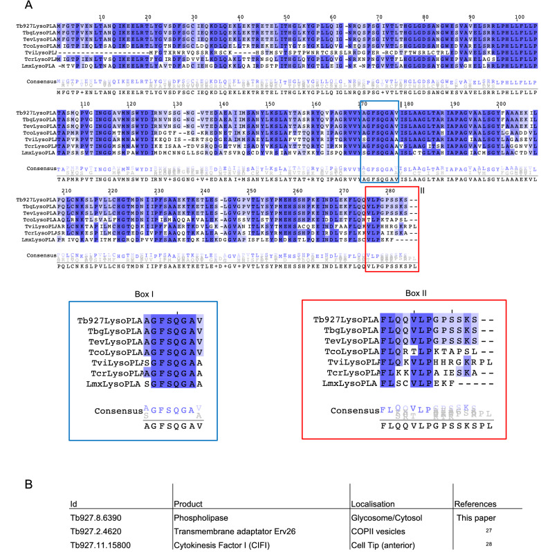
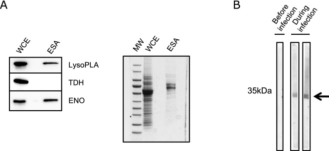
Phospholipases are esterases involved in lipid catabolism. In pathogenic micro-organisms (bacteria, fungi, parasites) they often play a critical role in virulence and pathogenicity. A few phospholipases (PL) have been characterised so far at the gene and protein level in unicellular parasites including African trypanosomes (AT). They could play a role in different processes such as host-pathogen interaction, antigenic variation, intermediary metabolism. By mining the genome database of AT we found putative new phospholipase candidate genes and here we provided biochemical evidence that one of these has lipolytic activity. This protein has a unique non-canonical glycosome targeting signal responsible for its dual localisation in the cytosol and the peroxisomes-related organelles named glycosomes. We also show that this new phospholipase is excreted by these pathogens and that antibodies directed against this protein are generated during an experimental infection with T. brucei gambiense, a subspecies responsible for infection in humans. This feature makes this protein a possible tool for diagnosis.


Conflict of interest statement

The authors declare no competing interests.

Figures

Figure 1
Figure 1
Comparison of LysoPLA in kinetoplastids. (A) Alignment of LysoPLA from kinetoplastids. Sequences were extracted and aligned using Clustal omega. Dark blue contains conserved residues, white to light blue contains conservative changes. Box I emphasizes the conservation of the putative phospholipase active site. Box II emphasizes the differences between kinetoplastids LysoPLA Carboxy-terminus. Tb, Trypanosoma brucei brucei (Tb927.8.6390); Tbg, Trypanosoma brucei gambiense (Tbg972.8.6450), Tev, Trypanosoma evansi (TevSTIB805.5.6680); Tco, Trypanosoma congolense (TcIL3000_8_6240); Tv, Trypanosoma vivax (TvY486_0805980); Tcr, Trypanosoma cruzi (TcCLB.506797.70); Lmx, Leishmania Mexicana (LmxM.24.1840). (B) Only 3 proteins are ending with SKS in Trypanosoma brucei brucei. The search was performed on TriTrypDB.org using the « Protein Motif Pattern » tool.
Figure 2
Figure 2
Lipolytic activity of recombinant TbLysoPLA. (A) PLA1 activity assay on recombinant proteins expressed in E. coli. Purification steps were analysed by coomassie gel (above gel). As examplified for TbLysoPLA, SF (Soluble Fraction), FT (Flow Through), W1-2 (Washes), CE1-2 (Clivage/Elution after thrombin release). Tested fractions were dialysed against PBS. PLA1 activity was measured using Enzcheck PLA1 assay (Molecular Probes). Control was a commercial phospholipase A1 (Sigma L3295). TbLysoPLA, full-length TbLysoPLA; TbLysoPLASA171, mutated TbLysoPLA where the putative active serine 171 was replaced by an alanine; GST, Glutathione-S-Transferase was eluted with 20 mM glutathione instead of thrombin cleavage. Supplemental information concerning expression and purification can be found on Fig. S2. (B) Substrate specificity of recombinant TbLysoPLA. TbLysoPLA was incubated (B) or not (A) with a Lipid Mix containing lyso-PC C17:0, PC (diC16:0), PC O-C16, 20:5, PC (diC18:0).
Figure 3
Figure 3
Analysis of bloodstream form mutants. (A) Left: TbLysoPLA is expressed in both bloodstream and procyclic forms. Total protein extracts from both forms of Tb were resolved by SDS-PAGE and transferred on nitrocellulose as described in materiel and method section. Membranes were probed with sera raised against TbLysoPLA and PFR. Right panel is a control with the pre-immune sera from the animal before immunisation with TbLysoPLA. Middle: inhibition of TbLysoPLA expression by interference RNA. Growth of the parental cell-line and 3 independant clones cultivated with (i) or without (ni) tetracycline. Right panel shows the ∆TbLysoPLA validation by Western Blotting as TbLysoPLA is not detected anymore. A specific affinity-purified anti-TbLysoPLA antibody was used and PFR was used as loading control. (B) Mice infection with wild-type (dark grey) and ∆TbLysoPLA (light grey) Tb427 BSF. Error bars represent the standard error of the mean (n = 4 per group). Left: parasitemia of infected mice quantified by hemocytometer. Right: number of parasites in heart, kidney and gonadal adipose tissue, 5 days after infection, quantified by qPCR.
Figure 4
Figure 4
Subcellular distribution of TbLysoPLA in bloodstream forms. Digitonin titration (left of B, C and D): Western blot analysis of the supernatant (s) and pellet (p) fractions from cells incubated with 0.01–0.4 mg digitonin/mg protein in STE buffer containing 150 mM NaCl. The digitonin concentration required to release cytosolic (cyt) and glycosomal (gly) marker proteins are indicated by vertical dash line. TbLysoPLA was detected with specific anti-TbLysoPLA in (A) and anti-Ty in (B), (C) and (D). For technical reasons two gels/membranes were needed to analyse the whole set of samples. The Vertical line between 100 and 160 indicates a delimitation between two different membranes. Hypotonic lysis (middle of B, C and D): Western blot analysis of supernatant (s) and pellet (p) fractions from cells incubated with hypotonic buffer. Anti-Ty was used for detection of TbLysoPLA. Immunofluorescence Assay (IFA; right of B, C and D). Cells were stained with monoclonal mouse anti-Ty (Fluorescein channel) and rabbit anti-Aldolase (Alexa 568 channel). Nucleus and kinetoplast are stained with DAPI. Anti-Ty was used for detection of TbLysoPLA.
Figure 5
Figure 5
TbLysoPLA as a potent excreted/secreted factor. (A) Left: detection of LysoPLA in whole cell extract (WCE) of WT Tb BSF and in excreted/secreted material antigens (ESA). Specific anti-TbLysoPLA was used for detection, enolase was the positive control and TDH was used as a negative control. Right: commassie stating showing WCE and ESA. (B) Antibodies directed against TbLysoPLA are produced during infection Recombinant full-length TbLysoPLA was resolved by SPS-PAGE followed by a Western Blot. Mice serum taken before and during infection by T.b gambiense were used as primary antibody. Signal is detected only during infection (arrow).

References

    1. Barrett MP, Boykin DW, Brun R, Tidwell RR. Human African trypanosomiasis: Pharmacological re-engagement with a neglected disease. Br. J. Pharmacol. 2007;152:1155–1171. - PMC - PubMed
    1. Van den Bossche P. Some general aspects of the distribution and epidemiology of bovine trypanosomosis in southern Africa. Int. J. Parasitol. 2001;31:592–598. - PubMed
    1. Dennis EA. 9 Phospholipases. In: Boyer PD, editor. The Enzymes. Elsevier; 1983. pp. 307–353.
    1. Aoki J, Inoue A, Makide K, Saiki N, Arai H. Structure and function of extracellular phospholipase A1 belonging to the pancreatic lipase gene family. Biochimie. 2007;89:197–204. - PubMed
    1. Burke JE, Dennis EA. Phospholipase A2 structure/function, mechanism, and signaling. J. Lipid Res. 2009;50(Suppl):S237–242. - PMC - PubMed

Publication types