. 2022 Mar 19;12(1):4759.
doi: 10.1038/s41598-022-07756-6.

End-point RT-PCR based on a conservation landscape for SARS-COV-2 detection

Collaborators, Affiliations

End-point RT-PCR based on a conservation landscape for SARS-COV-2 detection

Armando Cruz-Rangel et al. Sci Rep. .

Abstract

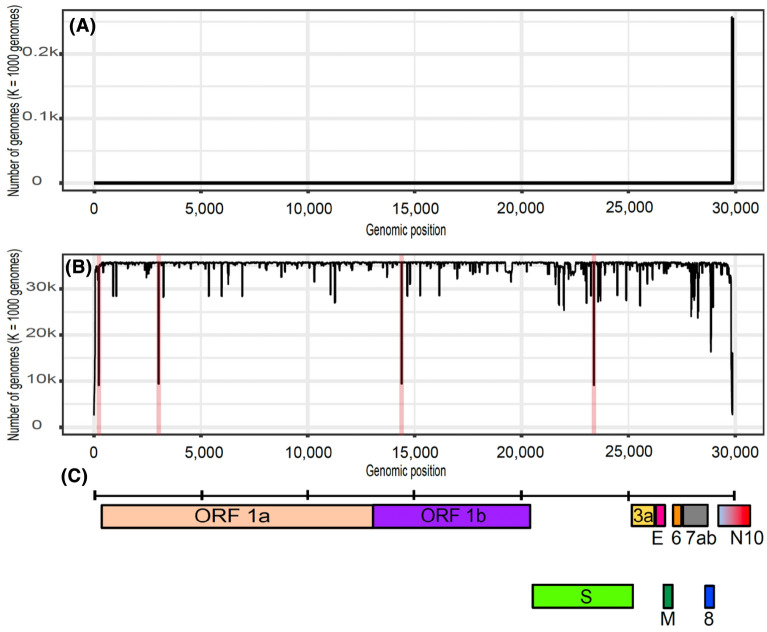
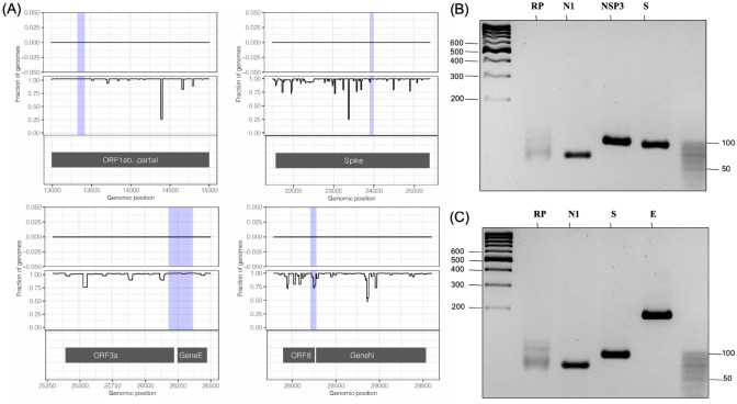
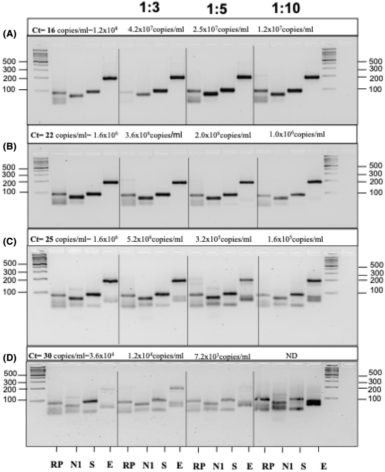
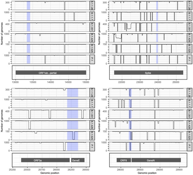
End-point RT-PCR is a suitable alternative diagnostic technique since it is cheaper than RT-qPCR tests and can be implemented on a massive scale in low- and middle-income countries. In this work, a bioinformatic approach to guide the design of PCR primers was developed, and an alternative diagnostic test based on end-point PCR was designed. End-point PCR primers were designed through conservation analysis based on kmer frequency in SARS-CoV-2 and human respiratory pathogen genomes. Highly conserved regions were identified for primer design, and the resulting PCR primers were used to amplify 871 nasopharyngeal human samples with a previous RT-qPCR based SARS-CoV-2 diagnosis. The diagnostic test showed high accuracy in identifying SARS-CoV-2-positive samples including B.1.1.7, P.1, B.1.427/B.1.429 and B.1.617.2/ AY samples with a detection limit of 7.2 viral copies/µL. In addition, this test could discern SARS-CoV-2 infection from other viral infections with COVID-19-like symptomatology. The designed end-point PCR diagnostic test to detect SARS-CoV-2 is a suitable alternative to RT-qPCR. Since the proposed bioinformatic approach can be easily applied in thousands of viral genomes and over highly divergent strains, it can be used as a PCR design tool as new SARS-CoV-2 variants emerge. Therefore, this end-point PCR test could be employed in epidemiological surveillance to detect new SARS-CoV-2 variants as they emerge and propagate.


Conflict of interest statement

The authors declare no competing interests.

Figures

Figure 1
Figure 1
(A) Genome-wide environmental conservation landscape. (B) Genome-wide population conservation landscape; positions 241, 3037, 14,408 and 23,403 are highlighted in red. In both panels, the X axis represents the genome position and the Y axis represents the number of genomes that contain each reference kmer. (C) Genome annotation.
Figure 2
Figure 2
(A) Environmental and population landscapes are shown for each selected region. A panel is included for all primers used in this study. The shadowed blue regions represent the amplicons generated by the PCR primers: upper left, primer NSP-3; upper right, primer S; lower left, primer E and lower right, primer N1. Upper track: number of occurrences of each reference kmer in the environmental sequences; upper and middle track: X-axis shows the genomic position for the start of each reference kmer, Y-axis, shows the percentage of either environmental sequences (upper track) or SARS-CoV-2 population genomes (middle track) that contain a given reference kmer; lower track, genomic annotation. (B) 4% agarose gels showing the end-point RT-PCR amplification product when using as sample a positive control (SARS-CoV-2 RNA) with the primers RP and N1 (CDC recommended primers) as well as the NSP3 and S primers generated by the kmer method. (C) 4% agarose gels showing the end-point RT-PCR amplification product when using as sample a positive control with the RP, N1, S and E primers.
Figure 3
Figure 3
End-point PCR showed a high detection limit when analyzing samples with a positive qRT-PCR diagnosis. All samples in the study had known Ct values that were used to determine viral copy numbers. (AD) Increasing Ct values in samples were correlated with lower viral loads. (D) The positive sample with a Ct value of 30 corresponded to a detection limit of 7 viral copies/µl (1:5 dilution). At lower dilutions, the virus fragments were no longer detectable, and nonspecific bands of human genomic bulk were observed.
Figure 4
Figure 4
Number of SARS-CoV-2 population genomes that contain each reference kmer. Each plot shows each genome position at the X axis and the number of occurrences of the kmer starting at position X at the Y axis for each group of SARS-CoV-2 genomes. The shadowed blue regions represent the regions amplified by the PCR primers used in this study: upper left, primer NSP-3; upper right, primer S; lower left, primer E and lower right, primer N1.
Figure 5
Figure 5
Assessment of the specificity of the SARS-CoV-2 detection test. (A) 4% agarose gel showing that the primers used for SARS-CoV-2 detection did not amplify H1N1 influenza virus RNA. (B) 4% agarose gel revealing that the test is adequate to discriminate SARS-CoV-2 from other coronaviruses such as β-coronavirus-OC43. M2, a specific fragment of ORF-7, and N1-OC43, a specific amplicon of the N gene, were used as positive controls for H1N1 influenza virus and β-coronavirus, respectively.

Similar articles

Cited by

References

    1. Wu F, et al. A new coronavirus associated with human respiratory disease in China. Nature. 2020;579:265–269. doi: 10.1038/s41586-020-2008-3. - DOI - PMC - PubMed
    1. World Health Organization. https://covid19.who.int/ (2021).
    1. Covid Investigation Team Clinical and virologic characteristics of the first 12 patients with coronavirus disease 2019 (COVID-19) in the United States. Nat. Med. 2020;26:861–868. doi: 10.1038/s41591-020-0877-5. - DOI - PubMed
    1. de Assis RR, et al. Analysis of SARS-CoV-2 antibodies in COVID-19 convalescent blood using a coronavirus antigen microarray. Nat. Commun. 2021;12:6. doi: 10.1038/s41467-020-20095-2. - DOI - PMC - PubMed
    1. Diao B, et al. Accuracy of a nucleocapsid protein antigen rapid test in the diagnosis of SARS-CoV-2 infection. Clin. Microbiol. Infect. 2021;27(289):e1–289. - PMC - PubMed

Publication types

Supplementary concepts