. 2022 Mar 19;12(1):4752.
doi: 10.1038/s41598-022-08903-9.

A role for the periplasmic adaptor protein AcrA in vetting substrate access to the RND efflux transporter AcrB

Affiliations

A role for the periplasmic adaptor protein AcrA in vetting substrate access to the RND efflux transporter AcrB

Ilyas Alav et al. Sci Rep. .

Abstract

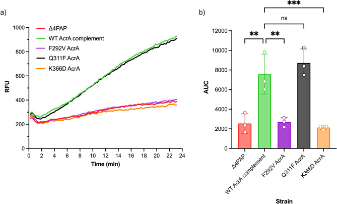
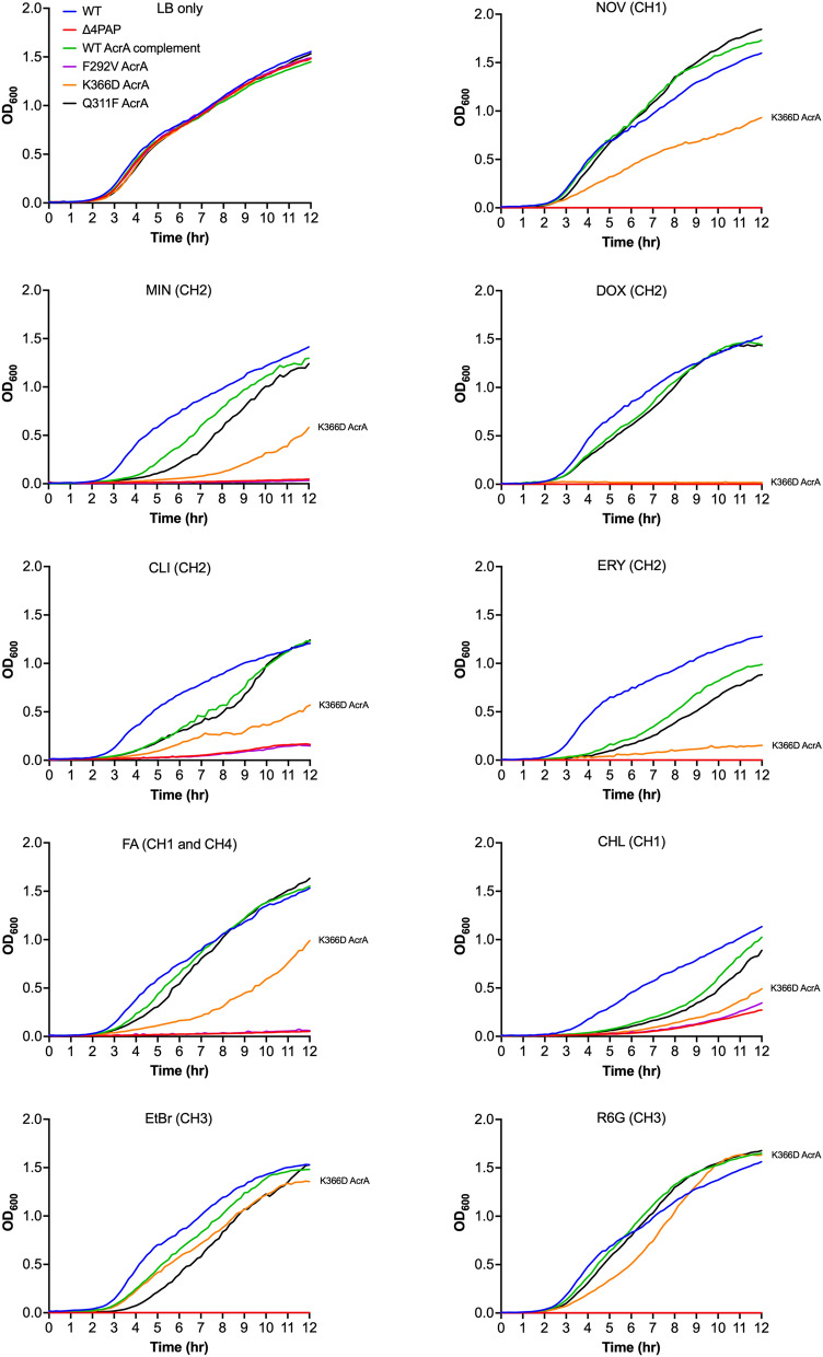
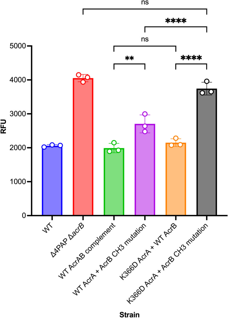
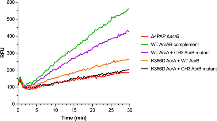
Tripartite resistance-nodulation-division (RND) efflux pumps, such as AcrAB-TolC of Salmonella Typhimurium, contribute to antibiotic resistance and comprise an inner membrane RND-transporter, an outer membrane factor, and a periplasmic adaptor protein (PAP). The role of the PAP in the assembly and active transport process remains poorly understood. Here, we identify the functionally critical residues involved in PAP-RND-transporter binding between AcrA and AcrB and show that the corresponding RND-binding residues in the closely related PAP AcrE, are also important for its interaction with AcrB. We also report a residue in the membrane-proximal domain of AcrA, that when mutated, differentially affects the transport of substrates utilising different AcrB efflux channels, namely channels 1 and 2. This supports a potential role for the PAP in sensing the substrate-occupied state of the proximal binding pocket of the transporter and substrate vetting. Understanding the PAP's role in the assembly and function of tripartite RND pumps can guide novel ways to inhibit their function to combat antibiotic resistance.


Conflict of interest statement

The authors declare no competing interests.

Figures

Figure 1
Figure 1
Multiple sequence alignment of Salmonella AcrA and AcrE combined with the mapping of the secondary structure derived from the experimentally defined structure of E. coli AcrA (PDB 5V5S, chain G). Identical residues are coloured red and similar residues are coloured yellow. The PAP-binding boxes implicated in RND-binding are numbered 1 to 9 and depicted using blue rectangles. Figure created using Espript 3.0.
Figure 2
Figure 2
Efflux of ethidium bromide by the Salmonella Typhimurium SL1344 Δ4PAP strain complemented with mutated versions of AcrA. Data presented are the mean of three biological replicates and are shown as the time taken for the fluorescence to decrease by 25% ± standard deviation. Bacteria were treated with ethidium bromide and the proton-motive force dissipator CCCP for 1 h and then re-energised with glucose. The annotation above indicates the mapping of each mutation to its binding box and the domain mapping of respective boxes. Data were analysed by one-way ANOVA and compared to the WT AcrA complement using Dunnett’s test. Strains with significantly different efflux are indicated with *** (P ≤ 0.001) or **** (P ≤ 0.0001).
Figure 3
Figure 3
Accumulation of ethidium bromide in the Salmonella Typhimurium SL1344 Δ4PAP ΔacrF strain complemented with mutated versions of AcrE. Data represented are the mean of three biological replicates showing maximum RFU values after 30 min of ethidium bromide exposure ± standard deviation. The annotation above indicates the mapping of each mutation to its binding box and the domain mapping of respective boxes. Data were analysed by one-way ANOVA and compared to the WT AcrE complement strain using Dunnett’s test. Strains with significantly different ethidium bromide accumulation are indicated with **** (P ≤ 0.0001). MPD, membrane-proximal domain.
Figure 4
Figure 4
(a) The crystal structure of the trimeric AcrB transporter and the hexameric AcrA assembly (TolC not shown for clarity). The different substrate entry pathways are shown as coloured channels, and the binding pockets are indicated by coloured spheres. (b) Zoomed-in view of the substrate channels and the binding pockets relative to K366 of AcrA. The green and orange spheres correspond to the space-fill representation of the residues lining the proximal binding pocket (PBP) and the deep binding pocket (DBP), respectively. K366 is in the membrane-proximal domain of AcrA and impacts the residues lining the PBP and the entrance of channel 2 (CH2). Channel 1 (CH1) also feeds into the PBP, so is likely to be impacted by changes in K366. Channel 3 (CH3) starts from the central cavity and leads to the DBP. Similarly, channel 4 (CH4) starts from the groove formed by TM1/TM2 and leads to the DBP. Therefore, CH3 and CH4 are unlikely to be directly impacted by K366 substitutions.
Figure 5
Figure 5
(a) Efflux of doxorubicin over time in the Salmonella Typhimurium SL1344 Δ4PAP strain complemented with mutated versions of AcrA. Bacteria were treated with doxorubicin and the proton-motive force dissipater CCCP for one hour and then re-energised with glucose. Efflux was monitored by increasing RFU due to extracellular doxorubicin. Data presented are the mean of three biological replicates. (b) Area under curve (AUC) analysis for doxorubicin efflux over time. The data shown are the mean AUC of the three biological replicates shown in (a) ± standard deviation. Data were analysed by one-way ANOVA and compared to the WT AcrA complement using Dunnett’s test. Strains with significantly different AUC are indicated with ** (P ≤ 0.01) or *** (P ≤ 0.001). ns, not significant.
Figure 6
Figure 6
Growth kinetics of the Salmonella Typhimurium SL1344 Δ4PAP strain complemented with mutated versions of AcrA. Abbreviations and concentrations of drugs used: CHL, 0.5 μg mL−1 chloramphenicol; CLI, 8 μg mL−1 clindamycin; DOX, 4 μg mL−1 doxorubicin; EtBr, 16 μg mL−1 ethidium bromide; ERY, 4 μg mL−1 erythromycin; FA, 8 μg mL−1 fusidic acid; MIN, 0.25 μg mL−1 minocycline; NOV, 4 μg mL−1 novobiocin; R6G, 16 μg mL−1 rhodamine 6G. Brackets indicate the preferred channel utilised by the substrate: CH1, channel 1; CH2, channel 2; CH3, channel 3; CH4, channel 4. The data shown are the mean OD600 values of three biological replicates. Concentrations of drugs are 0.25× MIC of K366D AcrA.
Figure 7
Figure 7
Accumulation of ethidium bromide in the Salmonella Typhimurium SL1344 Δ4PAP ΔacrB strain complemented with K366D AcrA and AcrB channel 3 (A33W T37W N298W AcrB) mutation. The data shown are the mean of three biological replicates showing maximum RFU values after 30 min of ethidium bromide exposure ± standard deviation. Data were analysed by one-way ANOVA and corrected for multiple comparisons using Tukey’s test. Strains with significantly different ethidium bromide accumulation are indicated with ** (P ≤ 0.01) or **** (P ≤ 0.0001). ns, not significant.
Figure 8
Figure 8
Efflux of doxorubicin over time in the Salmonella Typhimurium SL1344 Δ4PAP ΔacrB strain complemented with K366D AcrA and the AcrB channel 3 (CH3) mutation. Bacteria were treated with doxorubicin and the proton-motive force dissipater CCCP for 1 h and then re-energised with glucose. Efflux was monitored by increasing RFU due to extracellular doxorubicin. Data presented are the mean of three biological replicates. AcrB CH3 mutation refers to A33W T37W N298W AcrB.

References

    1. Ventola CL. The antibiotic resistance crisis: Part 1: causes and threats. Pharm. Ther. 2015;40:277–283. - PMC - PubMed
    1. Blair JM, Webber MA, Baylay AJ, Ogbolu DO, Piddock LJ. Molecular mechanisms of antibiotic resistance. Nat. Rev. Microbiol. 2015;13:42–51. - PubMed
    1. Du D, et al. Multidrug efflux pumps: Structure, function and regulation. Nat. Rev. Microbiol. 2018;16:523–539. - PubMed
    1. Colclough AL, et al. RND efflux pumps in Gram-negative bacteria; regulation, structure and role in antibiotic resistance. Future Microbiol. 2020;15:143–157. - PubMed
    1. Li XZ, Plesiat P, Nikaido H. The challenge of efflux-mediated antibiotic resistance in Gram-negative bacteria. Clin. Microbiol. Rev. 2015;28:337–418. - PMC - PubMed

Publication types

MeSH terms

Substances