

Practice Guideline
doi: 10.1016/j.jacc.2022.02.003.
Epub 2022 Mar 16.
2022 ACC Expert Consensus Decision Pathway on Cardiovascular Sequelae of COVID-19 in Adults: Myocarditis and Other Myocardial Involvement, Post-Acute Sequelae of SARS-CoV-2 Infection, and Return to Play: A Report of the American College of Cardiology Solution Set Oversight Committee
- PMID: 35307156
- PMCID: PMC8926109
- DOI: 10.1016/j.jacc.2022.02.003
Item in Clipboard
Practice Guideline
2022 ACC Expert Consensus Decision Pathway on Cardiovascular Sequelae of COVID-19 in Adults: Myocarditis and Other Myocardial Involvement, Post-Acute Sequelae of SARS-CoV-2 Infection, and Return to Play: A Report of the American College of Cardiology Solution Set Oversight Committee
J Am Coll Cardiol.
.
No abstract available
Keywords: ACC Expert Consensus Decision Pathway; COVID-19; SARS-CoV-2; athletes; cardiovascular diseases; chest pain; myocarditis; sports medicine; vaccines.
Figures
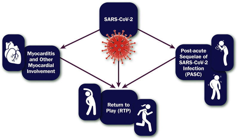
Scope of the Expert Consensus Decision Pathway on Cardiovascular Sequelae of COVID-19 in Adults PASC = post-acute sequelae of SARS-CoV-2 infection; RTP = return to play; SARS-CoV-2 = severe acute respiratory syndrome coronavirus 2.
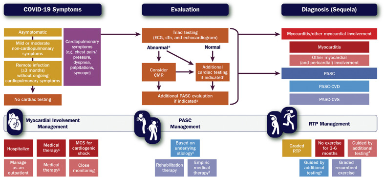
Framework for Evaluating and Managing Cardiovascular Sequelae of COVID-19 in Adults Gold and purple boxes = COVID-19 symptoms. Orange boxes = cardiac testing. Red and blue boxes = diagnoses (sequelae). ∗Includes elevated cTn; ECG with diffuse T-wave inversion, ST-segment elevation without reciprocal ST-segment depression, and/or prolongation of the QRS complex duration; and echocardiogram with ventricular wall motion abnormalities, often in a noncoronary distribution, and/or abnormal ventricular strain. †Informed by the presentation and may include coronary angiography for suspected acute coronary syndrome or CT pulmonary angiography for suspected pulmonary embolism (see Figure 3). ‡Includes other laboratory testing (eg, complete blood count, basic metabolic panel, C-reactive protein), an ambulatory rhythm monitor, a chest X-ray or CT imaging, and pulmonary function tests, along with additional testing for suspected PASC-CVD or PASC-CVS (see Figure 7). §For patients with myocarditis, medical therapy may include immunosuppressive drugs (eg, corticosteroids); for patients with pericardial involvement, medical therapy may include nonsteroidal anti-inflammatory drugs, colchicine, and corticosteroids (see Figure 3). ‖See Figure 7. ¶For patients with PASC-CVS, empiric medical therapy may include a beta-blocker, a non-dihydropyridine calcium-channel blocker, ivabradine, fludrocortisone, and/or midodrine. #Includes maximal-effort exercise testing and/or an ambulatory rhythm monitor (see Figure 9). CMR = cardiac magnetic resonance imaging; COVID-19 = novel coronavirus disease 2019; CT = computed tomography; cTn = cardiac troponin; ECG = electrocardiogram, MCS = mechanical circulatory support; PASC = post-acute sequelae of SARS-CoV-2 infection; PASC-CVD = PASC-Cardiovascular Disease; PASC-CVS = PASC-Cardiovascular Syndrome; RTP = return to play; SARS-CoV-2 = severe acute respiratory syndrome coronavirus 2.
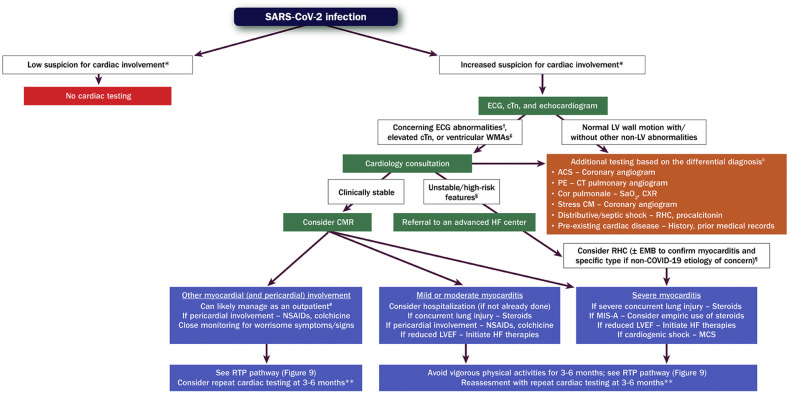
Evaluation and Management of Patients With Suspected Myocarditis or Myocardial Involvement Green boxes = cardiac testing for evaluation of myocarditis/myocardial involvement. Orange box = other cardiac/noncardiac testing. Purple boxes = management. ∗Informed by symptoms suggestive of cardiac involvement, including chest pain/pressure, dyspnea, palpitations, and syncope. †Includes diffuse T-wave inversion, ST-segment elevation without reciprocal ST-segment depression, and prolongation of the QRS complex duration. ‡Often in a noncoronary distribution; may also include abnormal ventricular strain. §Includes hypotension, cardiogenic shock, sustained ventricular arrhythmias, and/or advanced atrioventricular block. ‖This is an incomplete list of potential etiologies. ¶Testing for viral genomes should be performed on frozen heart tissue to exclude other causes of myocarditis, if possible. #Assumes chest pain is the only symptom, LV systolic function is preserved, and there are no ventricular arrhythmias. ∗∗Includes an ECG, echocardiogram, ambulatory rhythm monitor, and CMR. ACS = acute coronary syndrome; CM = cardiomyopathy; CMR = cardiac magnetic resonance imaging; COVID-19 = novel coronavirus disease 2019; CT = computed tomography; cTn = cardiac troponin; CXR = chest X-ray; ECG = electrocardiogram; EMB = endomyocardial biopsy; HF = heart failure; LV = left ventricular; LVEF = left ventricular ejection fraction; MCS = mechanical circulatory support; MIS-A = multisystem inflammatory syndrome in adults; NSAIDs = nonsteroidal anti-inflammatory drugs; PE = pulmonary embolism; RHC = right heart catheterization; SaO2 = arterial oxygen saturation; SARS-CoV-2 = severe acute respiratory syndrome coronavirus 2; WMAs = wall motion abnormalities.
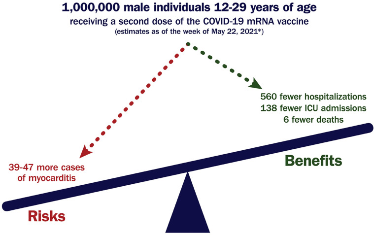
Favorable Benefit-to-Risk Ratio for COVID-19 mRNA Vaccination Among Those at Highest Risk for Postvaccination Myocarditis ∗Centers for Disease Control and Prevention. Coronavirus disease 2019 (COVID-19)-associated hospitalization surveillance network (COVID-NET)., COVID-19 = novel coronavirus disease 2019; ICU = intensive care unit, mRNA = messenger RNA.
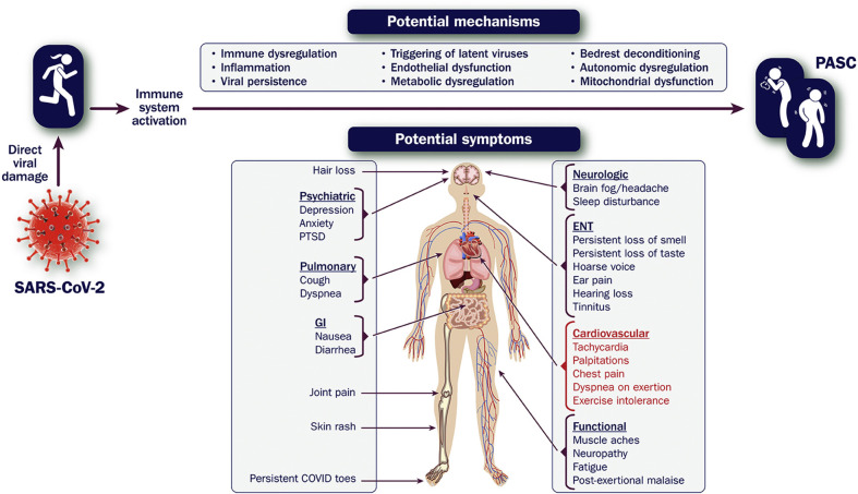
Symptoms of PASC and Potential Mechanisms COVID-19 = novel coronavirus disease 2019; ENT = ear, nose, and throat; GI = gastrointestinal; PASC = post-acute sequelae of SARS-CoV-2 infection; PTSD = posttraumatic stress disorder; SARS-CoV-2 = severe acute respiratory syndrome coronavirus 2.
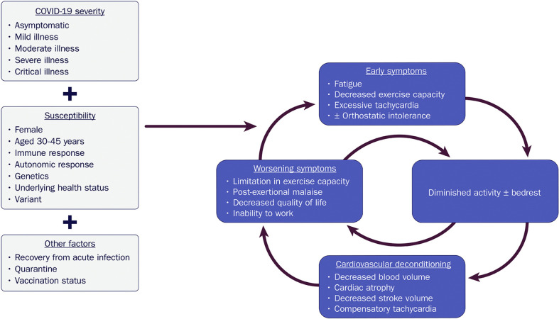
Downward Spiral of Deconditioning: A Potential Mechanism of Exercise Intolerance and Excessive Tachycardia in COVID-19 COVID-19 = novel coronavirus disease 2019.
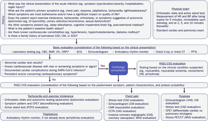
Evaluation of Cardiovascular Symptoms Suggestive of PASC ∗Consideration should be given to additional laboratory testing (eg, D-dimer, B-type natriuretic peptide/N-terminal pro-B-type natriuretic peptide, thyroid function tests) based on the clinical presentation. BMP = basic metabolic panel; BP = blood pressure; CAD = coronary artery disease; CBC = complete blood count; CCTA = coronary computed tomography angiography; CM = cardiomyopathy; CMR = cardiac magnetic resonance imaging; CPET = cardiopulmonary exercise test; CRP = C-reactive protein; CT = computed tomography; cTn = cardiac troponin; CV = cardiovascular; ECG = electrocardiogram; GI = gastrointestinal; HR = heart rate; MVD = microvascular dysfunction; PASC = post-acute sequelae of SARS-CoV-2 infection; PASC-CVD = PASC-Cardiovascular Disease; PASC-CVS = PASC-Cardiovascular Syndrome; PET = positron emission tomography; PFTs = pulmonary function tests; POTS = postural orthostatic tachycardia syndrome; SARS-CoV-2 = severe acute respiratory syndrome coronavirus 2; SCD = sudden cardiac death; VHD = valvular heart disease.
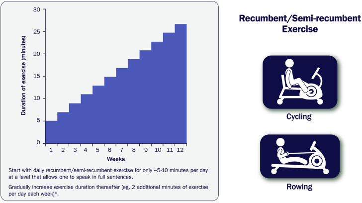
Sample Recumbent Exercise Therapy Prescription ∗For a more precise prescription for exercise therapy with heart rate targets and patient exercise logs, see appendix to Bryarly et al.
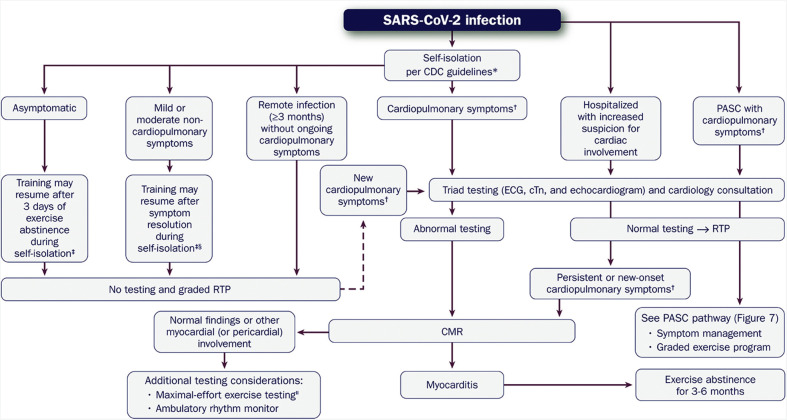
Evaluation of the Athletic Patient Convalesced From COVID-19 and Guidance on RTP and/or Intense Training ∗CDC Guidelines: COVID-19 Quarantine and Isolation. †Cardiopulmonary symptoms include chest pain/tightness, dyspnea, palpitations, and lightheadedness/syncope; this also includes symptoms occurring ≤1 week following COVID-19 mRNA vaccination. ‡Strategies to minimize transmission of SARS-CoV-2 to other athletes 3-10 days following a positive COVID-19 test include 1) training in isolation, 2) participating in socially-distanced outdoor training, 3) training with a face mask in a well-ventilated facility with appropriate social distancing, and 4) participating in group training after a single negative NAAT (eg, RT-PCR test) or 2 negative rapid antigen tests 24-48 hours apart. §Excludes prolonged, isolated anosmia/ageusia, which should not delay return to training. ‖Maximal-effort exercise testing should be deferred until myocarditis has been excluded. CDC = Centers for Disease Control and Prevention; CMR = cardiac magnetic resonance imaging; COVID-19 = novel coronavirus disease 2019, cTn = cardiac troponin; ECG = electrocardiogram; NAAT = nucleic acid amplification test, PASC = post-acute sequelae of SARS-CoV-2 infection; RTP = return to play; RT-PCR = reverse transcription polymerase chain reaction, SARS-CoV-2 = severe acute respiratory syndrome coronavirus 2.
References
-
- Januzzi J.L., Jr., Ahmad T., Binder L.G., et al. 2019 methodology for creating expert consensus decision pathways: a report of the American College of Cardiology. J Am Coll Cardiol. 2019;74:1138–1150. - PubMed
-
- Johns Hopkins University & Medicine Coronarvirus Resource Center https://coronavirus.jhu.edu/map.html
Publication types
MeSH terms
LinkOut - more resources
Full Text Sources
Medical
Miscellaneous
