Review
. 2022 Apr;600(8):1825-1837.
doi: 10.1113/JP281061. Epub 2022 Mar 30.

Hepatic stellate cells in physiology and pathology

Affiliations
Review

Hepatic stellate cells in physiology and pathology

Dakota R Kamm et al. J Physiol. 2022 Apr.

Abstract

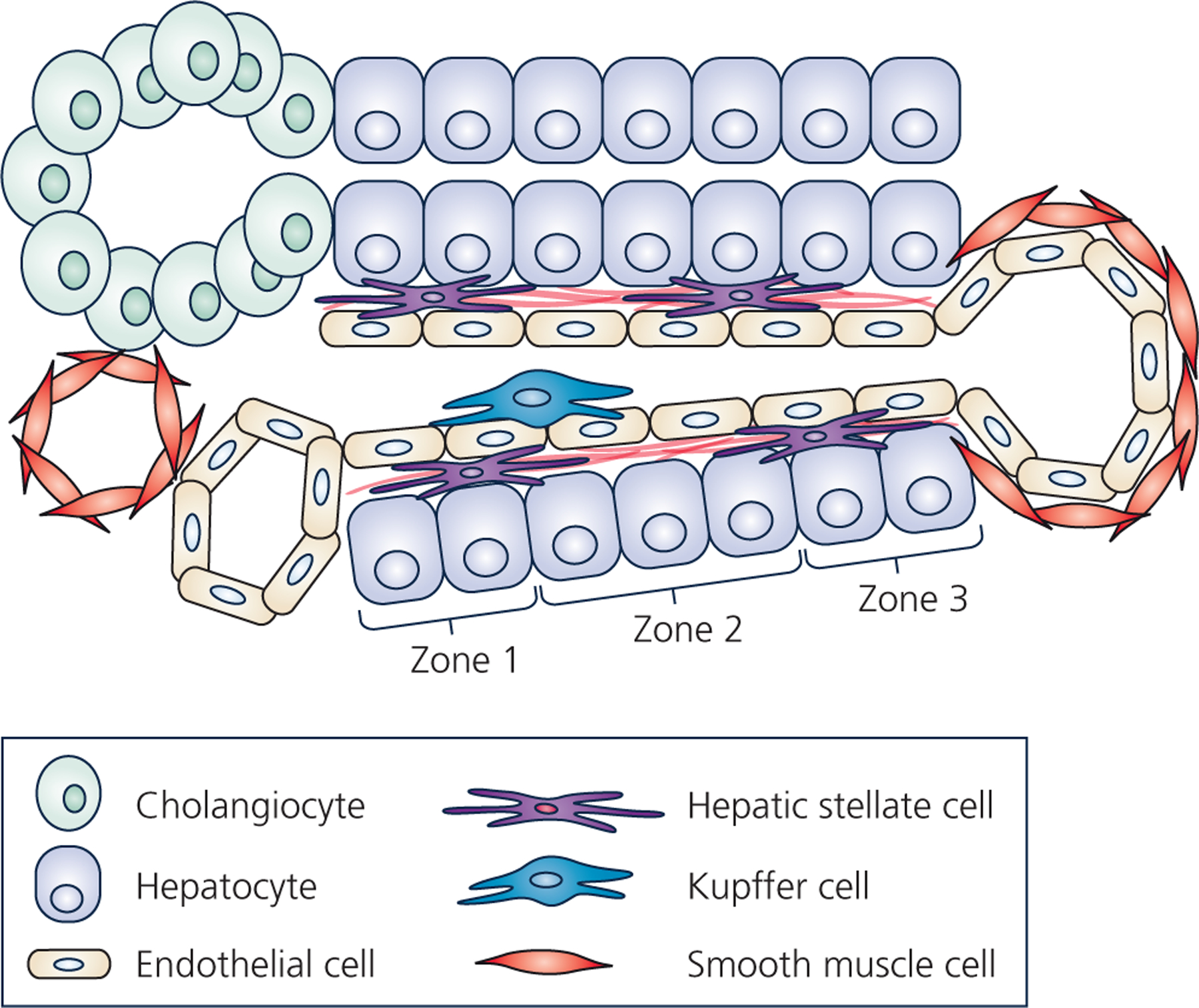
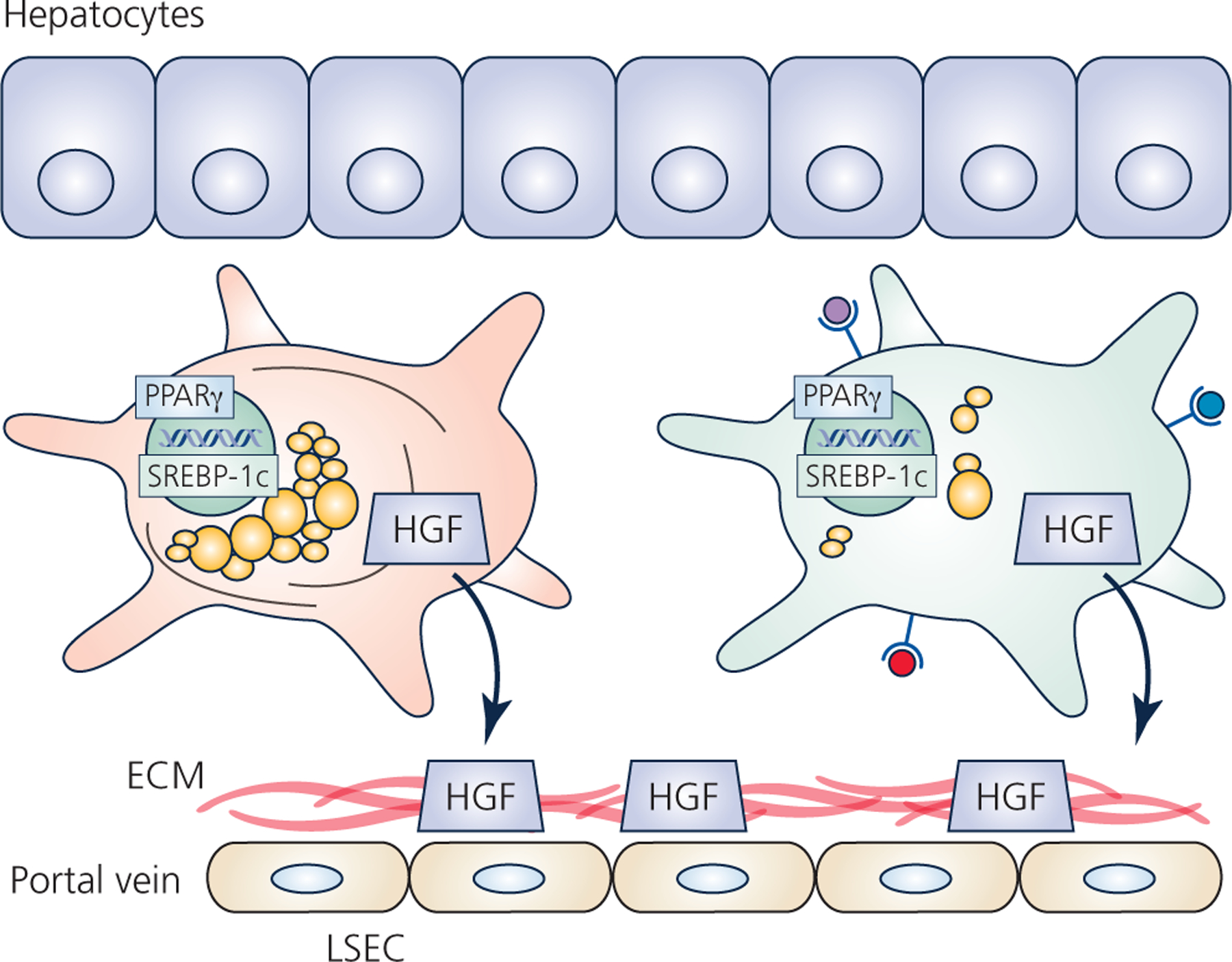
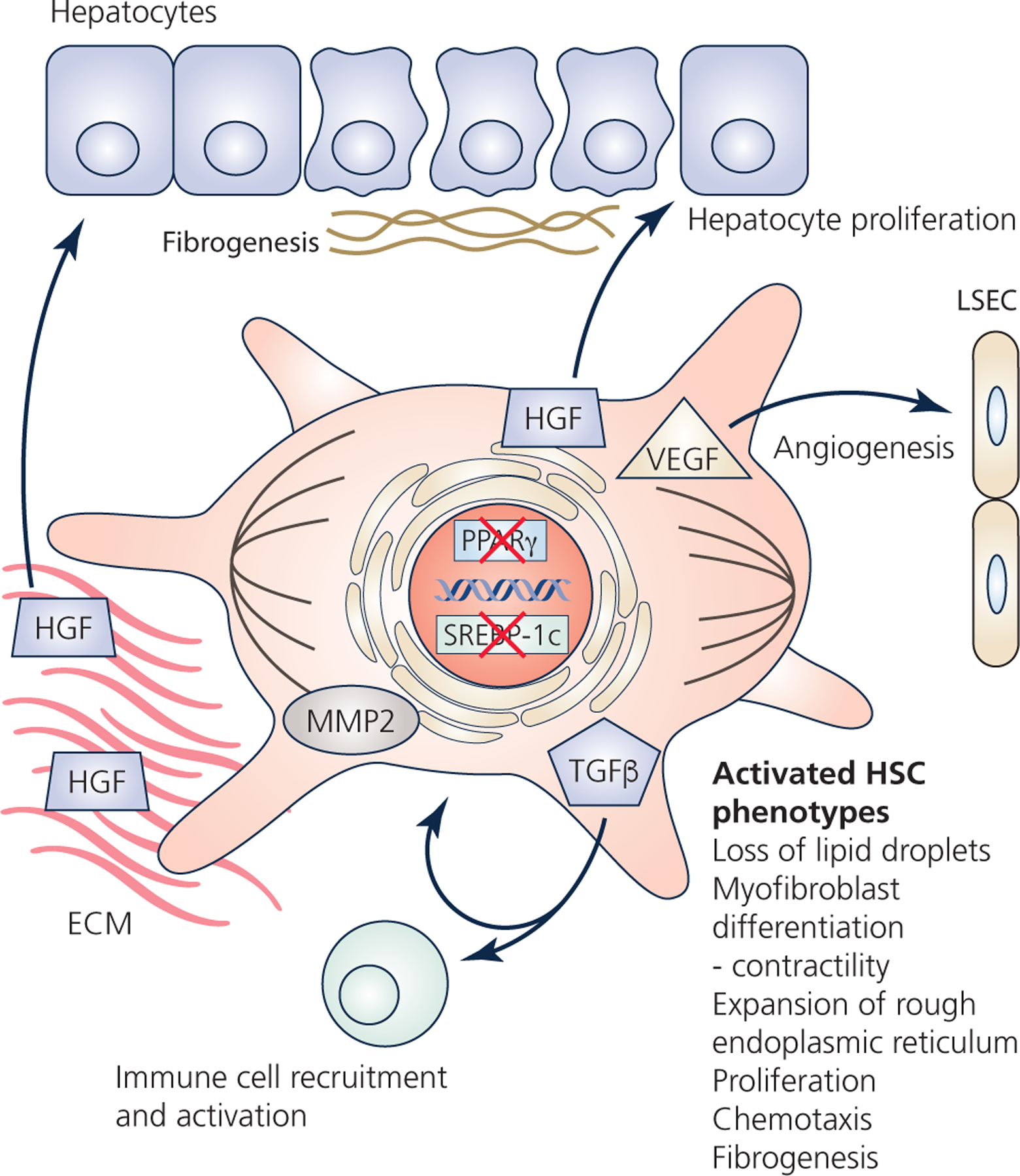
Hepatic stellate cells (HSCs) comprise a minor cell population in the liver but serve numerous critical functions in the normal liver and in response to injury. HSCs are primarily known for their activation upon liver injury and for producing the collagen-rich extracellular matrix in liver fibrosis. In the absence of liver injury, HSCs reside in a quiescent state, in which their main function appears to be the storage of retinoids or vitamin A-containing metabolites. Less appreciated functions of HSCs include amplifying the hepatic inflammatory response and expressing growth factors that are critical for liver development and both the initiation and termination of liver regeneration. Recent single-cell RNA sequencing studies have corroborated earlier studies indictaing that HSC activation involves a diverse array of phenotypic alterations and identified unique HSC populations. This review serves to highlight these many functions of HSCs, and to briefly describe the recent genetic tools that will help to thoroughly investigate the role of HSCs in hepatic physiology and pathology.

Keywords: fibrosis; liver; stellate cell.


Conflict of interest statement

Competing interests

The authors have no conflicts of interest to declare.

Figures

Figure 1 –
Figure 1 –. Hepatic cellular architecture.
Schematic represents a cross-section of a liver lobule, which contains the complete functional structure and main cell types of the liver. One the left, is the “portal triad” comprised of the portal vein, the hepatic artery, and the bile ductule. The bile ductule is comprised of cholangiocytes (represented as green oval-shaped cells), which collects bile produced by the hepatocytes. Bile ductules ultimately combine into the bile duct which drains bile to be stored in the gallbladder. The hepatic artery (represented as a red smooth-muscle cell-lined vessel) supplies oxygenated blood originating from the celiac artery, whereas the portal vein (represented as an endothelial cell-lined vessel) supplies nutrient- and toxin-rich blood from the stomach, pancreas, gallbladder, and spleen into the liver lobule. The portal vein and hepatic artery drain blood flow into the liver sinusoid, which is lined by liver sinusoidal endothelial cells. Ultimately, blood is collected into the central vein, lined with both endothelial cells and smooth muscle cells. Resident macrophages, known as Kupffer cells (represented as blue cell), reside in the luminal side of the sinusoid. The hepatic stellate cells (HSCs) (represented as peach-colored cells with long projections) reside in the space of Disse between hepatocytes and liver sinusoidal endothelial cells and produce the basement extracellular matrix (black lines near the HSCs in space of Disse). Regions along the length of the sinusoid are commonly referred to as zones, with zone 1 being peri-portal, zone 3 being peri-central vein, and zone 2 residing between. Hepatocytes comprise the hepatic parenchyma (the predominant cell type) and display an incredible array of functions including synthesis of serum proteins, clotting factors, lipoproteins, cholesterol and bile salts, gluconeogenesis and glycogen storage, as well as detoxification. Often these different hepatocyte functions are localized to specific zones along the sinusoid.
Figure 2 –
Figure 2 –. Functions and heterogeneity of quiescent HSCs.
All HSCs reside in the space of Disse between hepatocytes and liver sinusoidal endothelial cells (LSEC). Quiescence is maintained by high expression of the adipogenic transcription factors peroxisome proliferator-activated receptor gamma (PPARγ) and sterol regulatory-element-binding protein-1c (SREBP-1c). Quiescent HSCs also produce and secrete cytokines and growth factors, such as hepatocyte growth factor (HGF) which are sequestered by the basal extracellular matrix (ECM). Physically and functionally different HSCs likely reside in different regions along the portal tract. Early studies described distinct populations of HSCs based on presence/absence of desmin fibers, or abundance of lipid droplets. In addition to these macroscopic differences, single-cell RNA sequencing studies suggest other functional differences in these distinct populations, such as altered glycosaminoglycan synthesis or increased antigen presentation.
Figure 3 –
Figure 3 –. HSCs during liver injury and regeneration.
HSCs become activated by liver injury. Activation involves downregulation of the adipogenic transcription factors, and loss of the retinoid-storing lipid droplets. HSCs differentiate into contractile myofibroblasts, proliferate, and can respond to chemotactic signals. A number of secreted factors participate in the response to injury. For example, tumor growth factor beta (TGFβ) activates immune cells, but also enhances HSC activation via autocrine signaling. The HSCs continue to produce hepatocyte growth factor (HGF), but also degrade the basal extracellular matrix (ECM) by matrix metalloproteinase 2 (MMP2), which frees sequestered HGF and stimulates hepatocyte proliferation. HSCs also produce vascular endothelial growth factor (VEGF), as well as fibroblast growth factor, platelet-derived growth factor, and TGFβ which can all enhance angiogenesis. Activated HSCs also synthesize the collagen-rich fibrosis, as well as proteoglycans, glycosaminoglycans, and tissue-inhibitor of matrix metalloproteinases which all participate in the formation of the fibrotic scar. Lastly, HSCs participate in the termination of liver regeneration by re-establishing the basal ECM which sequesters HGF. Likewise, TGFβ from HSCs can inhibit hepatocyte proliferation to help terminate liver regeneration.

References

    1. Andrews TS, Atif J, Liu JC, Perciani CT, Ma XZ, Thoeni C, Slyper M, Eraslan G, Segerstolpe A, Manuel J, Chung S, Winter E, Cirlan I, Khuu N, Fischer S, Rozenblatt-Rosen O, Regev A, McGilvray ID, Bader GD & MacParland SA. (2021). Single-Cell, Single-Nucleus, and Spatial RNA Sequencing of the Human Liver Identifies Cholangiocyte and Mesenchymal Heterogeneity. Hepatol Commun. - PMC - PubMed
    1. Asahina K, Tsai SY, Li P, Ishii M, Maxson RE Jr., Sucov HM & Tsukamoto H (2009). Mesenchymal origin of hepatic stellate cells, submesothelial cells, and perivascular mesenchymal cells during mouse liver development. Hepatology 49, 998–1011. - PMC - PubMed
    1. Asahina K, Zhou B, Pu WT & Tsukamoto H. (2011). Septum transversum-derived mesothelium gives rise to hepatic stellate cells and perivascular mesenchymal cells in developing mouse liver. Hepatology 53, 983–995. - PMC - PubMed
    1. Baba S, Fujii H, Hirose T, Yasuchika K, Azuma H, Hoppo T, Naito M, Machimoto T & Ikai I. (2004). Commitment of bone marrow cells to hepatic stellate cells in mouse. J Hepatol 40, 255–260. - PubMed
    1. Ballardini G, Groff P, Badiali de Giorgi L, Schuppan D & Bianchi FB. (1994). Ito cell heterogeneity: desmin-negative Ito cells in normal rat liver. Hepatology 19, 440–446. - PubMed

Publication types

MeSH terms

Substances