. 2022 Mar 2:13:851089.
doi: 10.3389/fphar.2022.851089. eCollection 2022.

SLC35B4 Stabilizes c-MYC Protein by O-GlcNAcylation in HCC

Affiliations

SLC35B4 Stabilizes c-MYC Protein by O-GlcNAcylation in HCC

Tao Jiang et al. Front Pharmacol. .

Abstract

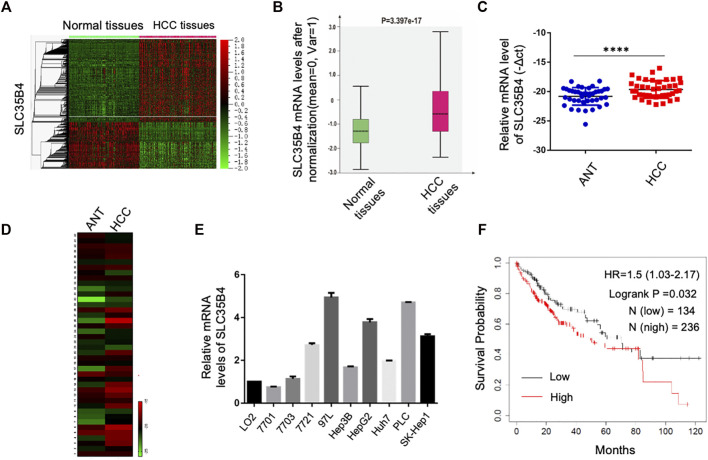
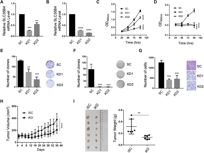
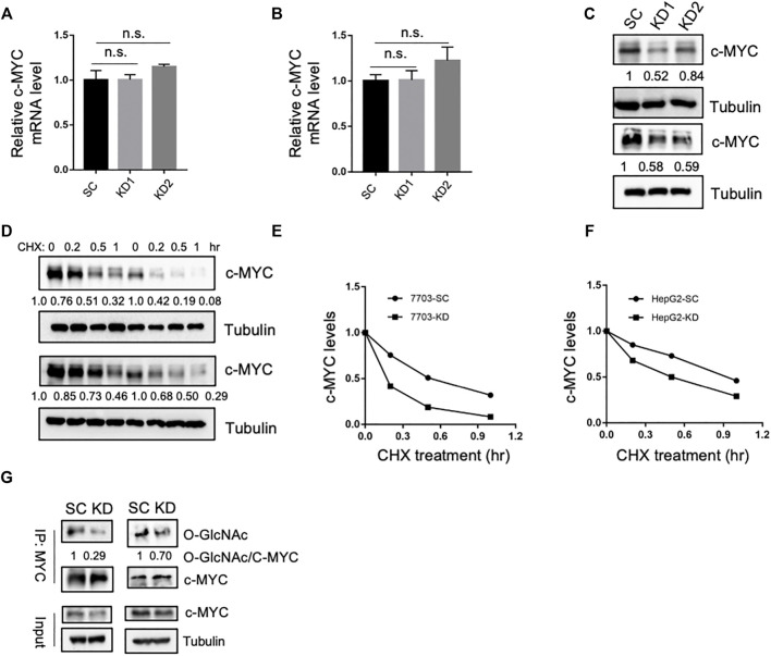
UDP-GlcNAc is a sugar substrate necessary for the O-GlcNAcylation of proteins. SLC35B4 is one of the nucleotide sugar transporters that transport UDP-GlcNAc and UDP-xylose into the endoplasmic reticulum and Golgi apparatus for glycosylation. The roles of SLC35B4 in hepatocellular carcinoma (HCC) tumorigenesis remain unknown. We find that the expression levels of SLC35B4 are higher in HCC tissues than adjacent non-tumor tissues. SLC35B4 is important for the proliferation and tumorigenesis of HCC cells. Mechanistically, SLC35B4 is important for the O-GlcNAc modification of c-Myc and thus the stabilization of c-Myc, which is required for HCC tumorigenesis. Therefore, SLC35B4 is a promising therapeutic target for treating HCC.

Keywords: HCC; O-GlcNAcylation; SLC35B4; c-Myc; nucleotide sugar transporters.


Conflict of interest statement

The authors declare that the research was conducted in the absence of any commercial or financial relationships that could be construed as a potential conflict of interest.

Figures

FIGURE 1
FIGURE 1
SLC35B4 is overexpressed in HCC and inversely correlated with prognosis of HCC patients. (A) The heat map of mRNA expression profile in 268 non-tumor adjacent tissues and 243 HCC tissues in GSE25097 dataset. The mRNA expression level of SLC35B4 was indicated by a white line. (B) The box plot of SLC35B4 expression levels in non-tumor tissues (268) and HCC tissues (243) in GSE25097 dataset. P-value is indicated. (C,D) The relative mRNA expression levels of SLC35B4 in 42 paired adjacent normal tissues (ANT) and HCC tissues. P-value is indicated. (E) The mRNA expression levels of SLC35B4 in HCC cell lines. N = 3. Data are presented as mean ± SD. (F) Kaplan–Meier survival curve of the overall survival of HCC patients with high and low SLC35B4 expression levels. The SLC35B4 expression levels were inversely correlated with the overall survival of HCC patients. P-value is indicated.
FIGURE 2
FIGURE 2
SLC35B4 promotes HCC cell proliferation and migration. (A,B) Relative mRNA expression levels of SLC35B4 in HCC cells after SLC35B4 knockdown. N = 3. Data are presented as mean ± SD. (C,D) SLC35B4 knockdown significantly decreased the cellular proliferation of HCC cells as determined by CCK8. N = 3. Data are presented as mean ± SD. (E,F) SLC35B4 knockdown dramatically decreased the clonal formation ability of 7703 (E) and HepG2 (F) cells. N = 3. Data are presented as mean ± SD. (G) SLC35B4 knockdown inhibited the migration of HCC cells in vitro. N = 3. Data are presented as mean ± SD. (H) The volume of xenograft tumors after the inducible knockdown of SLC35B4. iSC, inducible scrambled control; iKD, inducible knockdown. (I) The image and weight of xenograft tumors formed by HCC cells with or without inducible SLC35B4 knockdown in NSG mice 37 days after inducible knockdown. N = 6. Data are presented as mean ± SD. *p < 0.05, **p < 0.01, ***p < 0.001, ****p < 0.0001.
FIGURE 3
FIGURE 3
SLC35B4 stabilizes c-Myc via O-GlcNAc modification. (A,B) Relative mRNA expression levels of c-Myc in 7703 (A) and HepG2 (B) cells with SLC35B4 knockdown. N = 3. Data are presented as mean ± SD. n.s., non-significant. (C) The protein levels of c-Myc in 7703 (upper) and HepG2 (bottom) cells with SLC35B4 knockdown. The relative protein levels are indicated. (D) The protein levels of c-Myc protein in 7703 (upper) and HepG2 (bottom) cells after CHX treatment. (E,F) Quantification of the protein levels of c-Myc in 7703 (E) and HepG2 (F) cells with or without SLC35B4 knockdown at different time points after CHX treatment. The relative protein levels are indicated. (G) The O-GlcNAcylation of c-Myc after SLC35B4 knockdown. c-Myc in 7703 (left) and HepG2 (right) cells with or without SLC35B4 knockdown was immunoprecipitated and the levels of the O-GlcNAcylation were detected with anti-O-GlcNAc antibody. The relative levels of O-GlcNAcylated versus total c-Myc are indicated.

References

    1. Ashikov A., Routier F., Fuhlrott J., Helmus Y., Wild M., Gerardy-Schahn R., et al. (2005). The Human Solute Carrier Gene SLC35B4 Encodes a Bifunctional Nucleotide Sugar Transporter with Specificity for UDP-Xylose and UDP-N-Acetylglucosamine. J. Biol. Chem. 280, 27230–27235. 10.1074/jbc.M504783200 - DOI - PubMed
    1. Bacigalupa Z. A., Bhadiadra C. H., Reginato M. J. (2018). O-GlcNAcylation: Key Regulator of Glycolytic Pathways. J. Bioenerg. Biomembr 50, 189–198. 10.1007/s10863-018-9742-3 - DOI - PMC - PubMed
    1. Bazan B., Wiktor M., Maszczak-Seneczko D., Olczak T., Kaczmarek B., Olczak M. (2018). Lysine at Position 329 within a C-Terminal Dilysine Motif Is Crucial for the ER Localization of Human SLC35B4. PLoS One 13, e0207521. 10.1371/journal.pone.0207521 - DOI - PMC - PubMed
    1. Carabet L. A., Rennie P. S., Cherkasov A. (2018). Therapeutic Inhibition of Myc in Cancer. Structural Bases and Computer-Aided Drug Discovery Approaches. Int. J. Mol. Sci. 20, 120. 10.3390/ijms20010120 - DOI - PMC - PubMed
    1. Dang C. V. (2012). MYC on the Path to Cancer. Cell 149, 22–35. 10.1016/j.cell.2012.03.003 - DOI - PMC - PubMed