. 2022 Mar 3:13:852802.
doi: 10.3389/fphar.2022.852802. eCollection 2022.

Tofacitinib Decreases Autophagy of Fibroblast-Like Synoviocytes From Rheumatoid Arthritis Patients

Affiliations

Tofacitinib Decreases Autophagy of Fibroblast-Like Synoviocytes From Rheumatoid Arthritis Patients

M Vomero et al. Front Pharmacol. .

Abstract

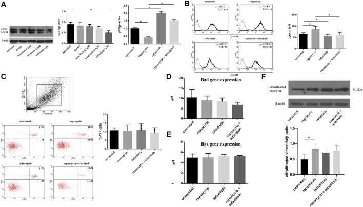
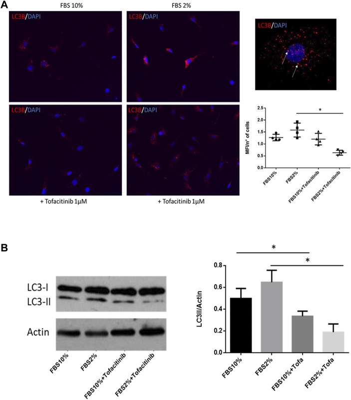
The pathway of Janus tyrosine kinases (JAKs) has a central role in the pathogenesis of Rheumatoid Arthritis (RA) by regulating multiple immune functions and cytokine production. The JAK inhibitor tofacitinib is effective in RA patients not responding to methotrexate or TNF-inhibitors. Since hyperactive autophagy has been associated with impaired apoptosis of RA fibroblast-like synoviocytes (FLS), we aimed to investigate the role of tofacitinib in modulating autophagy and apoptosis in these cells. FLS isolated from RA biopsies were cultured with tofacitinib in presence of autophagy inducer rapamycin and in serum deprivation condition. Levels of autophagy, apoptosis, and citrullinated proteins were analyzed by western blot, flow cytometry, immunocytofluorescence, and Real-Time PCR. Rapamycin induced an increase in RA-FLS autophagy while the levels of autophagy marker LC3-II were reduced after in vitro treatment with tofacitinib. The analysis of autophagic flux by specific fluorescence dye confirmed the reduction of autophagy in RA FLS. The treatment with tofacitinib did not influence apoptosis of RA FLS. Modulation of the autophagic process by tofacitinib did not significantly change citrullination. The results of this study demonstrate that tofacitinib is able to modulate autophagy of FLS contributing to its effectiveness in RA patients.

Keywords: Rheumatoid arthritis; apoptosis; autophagy; janus tyrosine kinases; tofacitinib.


Conflict of interest statement

The authors declare that the research was conducted in the absence of any commercial or financial relationships that could be construed as a potential conflict of interest.

Figures

FIGURE 1
FIGURE 1
Autophagy, apoptosis and citrullinated proteins levels after in vitro treatment with tofacitinib in FLS isolated from RA patients. (A). Western blot analysis of LC3-II and p62 levels of RA-FLS treated with tofacitinib (1uM) alone and in combination with rapamycin (200 nM). Blots shown are representative of five independent experiments performed using FLS samples from five different RA patients. Densitometry analysis of LC3-II and p62 levels relative to β-actin is also reported. Values are expressed as means ± sd. *p < 0.05 (B). Analysis of autophagosomes number in FLS treated with tofacitinib as previously described (n = 5). Values of mean fluorescence intensity (MFI) obtained by flow cytometry are reported. *p < 0.05 (C). Statistical analysis of apoptosis of FLS isolated from patients with RA (n = 5) after in vitro treatment with tofacitinib. Results are expressed as AV-positive cells. Dot plots (PI on y axis vs. AV on x axis) representative of five independent experiments are also shown. (D, E). Bad and Bax expressions of FLS treated with rapamycin (200 nM for 4 h) and tofacitinib (1 uM for 20 h) analyzed by Quantitative Real-time PCR (qRT-PCR). Data are reported as differences between their Ct and the housekeeping gene GAPDH Ct (ΔCt). (F). Western blot and densitometry analysis of citrullinated proteins levels in FLS from RA patients cultured in the presence of tofacitinib/rapamycin alone or in combination. Figure was chosen as representative of those obtained from five independent experiments on FLS from different RA patients. *p = 0.02.
FIGURE 2
FIGURE 2
Effect of tofacitinib on autophagy in FLS cultured in serum deprivation condition. (A) Immunocytofluorescence analysis of autophagy in FLS treated with tofacitinib alone and in starvation (FBS 2%). Autophagic LC3B + vesicles are stained in red in RA FLS treated with tofacitinib alone and in serum deprivation condition for 20 h (2% FBS). Cells were counterstained with DAPI dye to reveal DNA (blue staining). Autophagy is expressed as MFI normalized on the number of cells. The quantification was performed on four different areas for each condition. Images are representative of five independent experiments and higher magnification (×40) picture is also showed. *p < 0.05. (B) Western blot analysis of autophagy levels in FLS cultured in the presence of tofacitinib. Starvation was used to induce autophagy (FBS 2% for 20 h), and tofacitinib was used as a treatment for 20 h (1 µM). Autophagy was quantified by densitometric analysis on LC3-II and then normalized on β-Actin. Data are referred to five independent experiments in cells from different RA patients. *p < 0.05.

References

    1. Ando S., Kawada J. I., Watanabe T., Suzuki M., Sato Y., Torii Y., et al. (2016). Tofacitinib Induces G1 Cell-Cycle Arrest and Inhibits Tumor Growth in Epstein-Barr Virus-Associated T and Natural Killer Cell Lymphoma Cells. Oncotarget 7, 76793–76805. 10.18632/oncotarget.12529 - DOI - PMC - PubMed
    1. Colafrancesco S., Vomero M., Iannizzotto V., Minniti A., Barbati C., Arienzo F., et al. (2020). Autophagy Occurs in Lymphocytes Infiltrating Sjögren's Syndrome Minor Salivary Glands and Correlates with Histological Severity of Salivary Gland Lesions. Arthritis Res. Ther. 22, 238. 10.1186/s13075-020-02317-6 - DOI - PMC - PubMed
    1. Connor A. M., Mahomed N., Gandhi R., Keystone E. C., Berger S. A. (2012). TNFα Modulates Protein Degradation Pathways in Rheumatoid Arthritis Synovial Fibroblasts. Arthritis Res. Ther. 14, R62. 10.1186/ar3778 - DOI - PMC - PubMed
    1. Ichimura Y., Kominami E., Tanaka K., Komatsu M. (2008). Selective Turnover of p62/A170/SQSTM1 by Autophagy. Autophagy 4, 1063–1066. 10.4161/auto.6826 - DOI - PubMed
    1. Ireland J. M., Unanue E. R. (2011). Autophagy in Antigen-Presenting Cells Results in Presentation of Citrullinated Peptides to CD4 T Cells. J. Exp. Med. 208, 2625–2632. 10.1084/jem.20110640 - DOI - PMC - PubMed

LinkOut - more resources