Review
. 2022 Mar 3:13:852176.
doi: 10.3389/fmicb.2022.852176. eCollection 2022.

Force-Generation by the Trans-Envelope Tol-Pal System

Affiliations
Review

Force-Generation by the Trans-Envelope Tol-Pal System

Melissa N Webby et al. Front Microbiol. .

Abstract

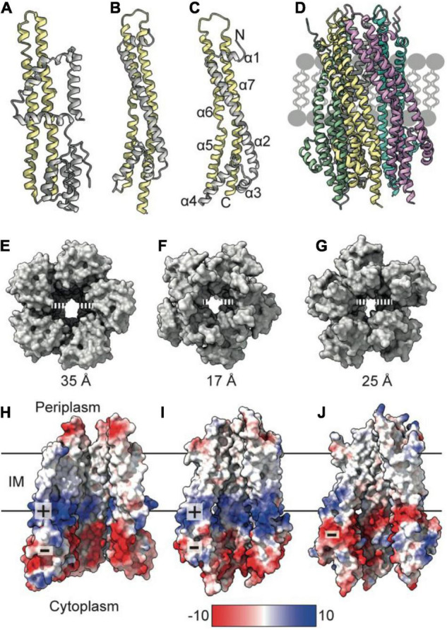
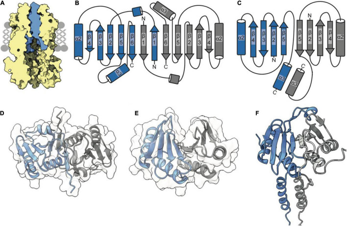
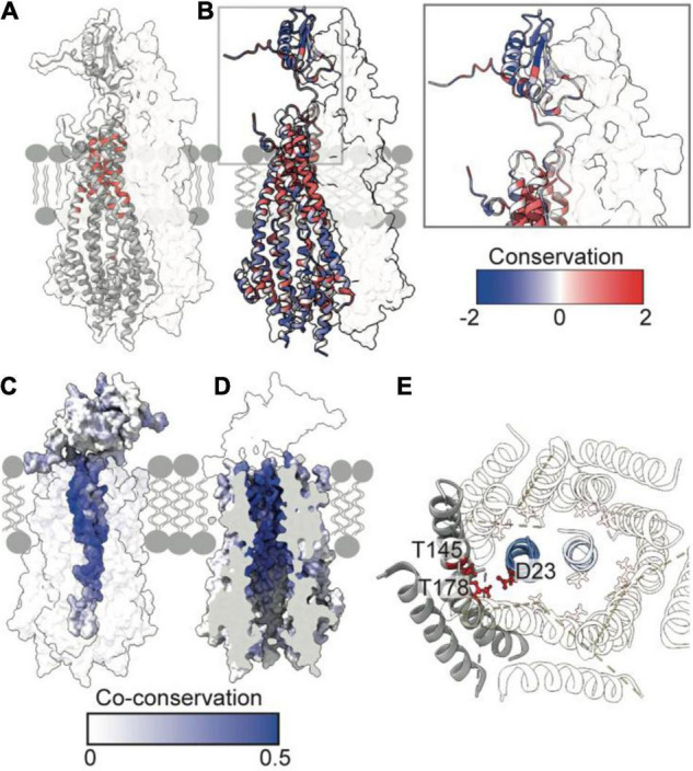
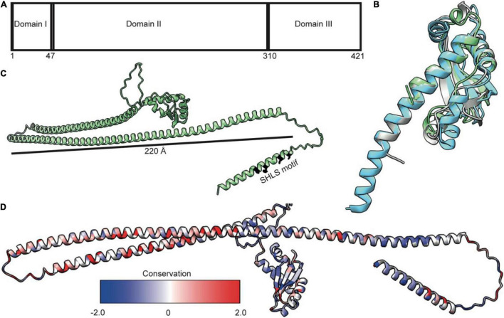
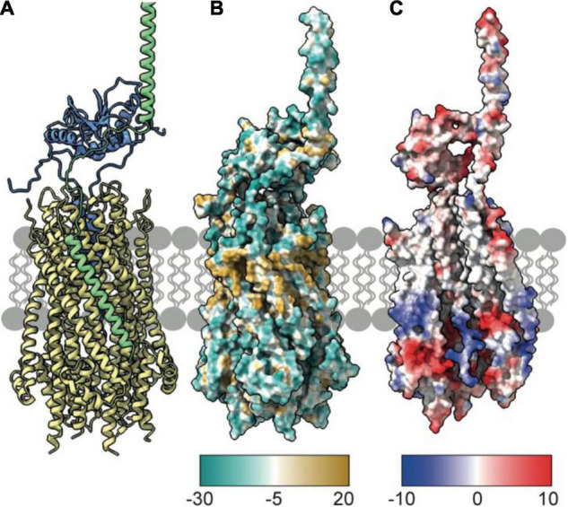
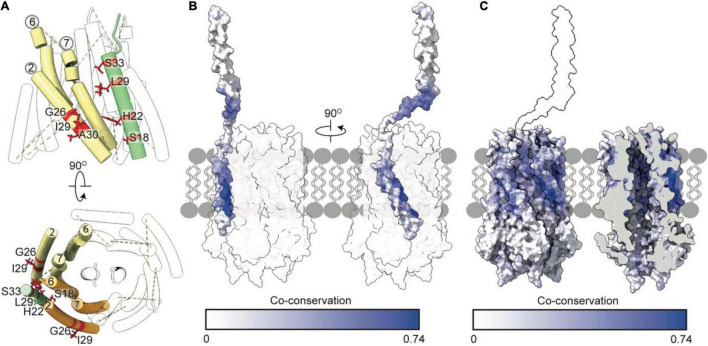
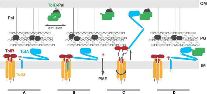
The Tol-Pal system spans the cell envelope of Gram-negative bacteria, transducing the potential energy of the proton motive force (PMF) into dissociation of the TolB-Pal complex at the outer membrane (OM), freeing the lipoprotein Pal to bind the cell wall. The primary physiological role of Tol-Pal is to maintain OM integrity during cell division through accumulation of Pal molecules at division septa. How the protein complex couples the PMF at the inner membrane into work at the OM is unknown. The effectiveness of this trans-envelope energy transduction system is underscored by the fact that bacteriocins and bacteriophages co-opt Tol-Pal as part of their import/infection mechanisms. Mechanistic understanding of this process has been hindered by a lack of structural data for the inner membrane TolQ-TolR stator, of its complexes with peptidoglycan (PG) and TolA, and of how these elements combined power events at the OM. Recent studies on the homologous stators of Ton and Mot provide a starting point for understanding how Tol-Pal works. Here, we combine ab initio protein modeling with previous structural data on sub-complexes of Tol-Pal as well as mutagenesis, crosslinking, co-conservation analysis and functional data. Through this composite pooling of in silico, in vitro, and in vivo data, we propose a mechanism for force generation in which PMF-driven rotary motion within the stator drives conformational transitions within a long TolA helical hairpin domain, enabling it to reach the TolB-Pal complex at the OM.

Keywords: Gram-negative bacteria; Tol-Pal; cell envelope; force transduction; outer membrane; proton motive force.


Conflict of interest statement

The authors declare that the research was conducted in the absence of any commercial or financial relationships that could be construed as a potential conflict of interest.

Figures

FIGURE 1
FIGURE 1
TolQ forms a pentameric pore. (A) MotA monomer transmembrane helices pack together to form a pentameric pore (yellow) and accessory helices are located on the exterior of the pore (gray). (B) ExbB monomer colored as in panel (A). (C) The TolQ monomer (E. coli AlphaFold model) colored as in panel (A), adopts a structure like that observed for ExbB. (D) Pentameric TolQ complex was generated following symmetry expansion of the AlphaFold monomer and energy minimization using RosettaRelax. Each monomer is colored separately. (E–G) The MotA, ExbB, and TolQ pentamers viewed from the cytoplasm to show the outward movement of helices that generate an expanded pore. By contrast, the structures can be overlaid almost perfectly at the periplasmic side. (H–J) A cut through the electrostatic surface of the MotA, ExbB, and TolQ pentamers, respectively, showing that the cytoplasmic side of the pore has a region of negative charge that is conserved across all three systems. A band of positive charge that spans the center of the pore is only observed in MotA and ExbB.
FIGURE 2
FIGURE 2
The TolR dimer has two distinct conformations. (A) The periplasmic entrance to the TolQ (yellow) pore is blocked by the transpore helices of the TolR dimer (blue). The TolQ-TolR model shown was assembled following alignment of the AlphaFold TolR monomer with the truncated TolR dimer from H. influenzae (TolRH). The full-length TolR dimer was then docked into the TolQ pentamer previously generated (Figure 1) based on position of ExbD in the ExbBD complex structure (Celia et al., 2019). The final TolQ-TolR complex shown was determined following energy minimization using RosettaRelax. (B) A depiction of the secondary structure of TolR from E. coli (TolRE) highlighting the strand-swapped dimer formed by β1 and β6 of each monomer. (C) A depiction of the secondary structure of TolRH has a dimer interface formed through interactions of β5-β5. (D) The TolRE strand-swapped dimer (PDB ID 5BY4) solved by X-ray crystallography shows the 180° rotation of one monomer (gray) relative to the other (blue). (E) The TolRH structure solved by NMR (PDB ID 2JWK) is in an open PG-binding conformation. Each monomer is represented in blue and gray, respectively. (F) The E. coli TolR dimer, generated in Rosetta, models the entirety of the protein, which adopts an open conformation like that of TolRH.
FIGURE 3
FIGURE 3
Essential, conserved, and co-conserved stator residues map to the TolQR lumen. (A) Red identifies essential residues that confer membrane instability when substituted (Vianney et al., 1994; Goemaere et al., 2007; Zhang et al., 2011). Two TolQ monomers are shown in cartoon format with a single TolR monomer. (B) The relative conservation of TolQ-TolR residues, which was generated following alignment (Clustal Omega) of ten TolQ sequences from different species, indicates that the most conserved residues map to the top or bottom of the TolQ lumen. The most-conserved residues of TolR map to the transpore helix and β-strand two. (C,D) The TolQ-TolR co-conservation profile of individual residues as scored by RaptorX suggest that the TolR helix inserts into the center of the TolQ pore. Co-conserved TolR and TolQ residues are predominantly within the transpore helix and lumen, respectively. (E) A ring of highly conserved threonine residues (red sticks) within the transmembrane region of TolQ (T145 in helix 6 and T178 in helix 7) are proposed to stabilize protonated TolR D23 (red sticks), based on structures of the MotA-MotB and ExbB-ExbD complexes (Celia et al., 2019; Deme et al., 2020).
FIGURE 4
FIGURE 4
TolA is a largely helical protein with three distinct domains. (A) TolA contains three annotated domains: domain I is the transmembrane helix, domain II is an elongated helical hairpin, and domain III is a small globular protein. Numbering is based on the E. coli sequence. (B) Structural overlay of TolA III from P. aeruginosa (cyan, PDB ID 1LR0) and V. cholerae (gray, PDB ID 4G7X) with the equivalent domain from E. coli (green, PDB ID 1S62) (RMSD of 1.3 and 2.0, respectively) showing that, although poorly conserved (20 and 25%, respectively), the domains have near identical folds. (C) The AlphaFold model of TolA from E. coli. The central domain II is predicted to form a helical hairpin that is 220 Å in length, which is long enough to span the periplasm. (D) Sequences of nine TolA proteins from different species were aligned in Clustal Omega, and sequence conservation was mapped onto the E. coli TolA structure within ChimeraX. Conserved residues are largely located within the C-terminal end of the helical hairpin of domain II. Residues in domains I and III are less conserved.
FIGURE 5
FIGURE 5
TolA packs against the TolQ pentamer to form the TolQRA complex. (A) The Rosetta model of the TolQ-TolR-TolA complex from E. coli was generated following placement of full length TolA, taken from the AlphaFold database, within 10 Å of the TolQ-TolR complex (Figure 2). A series of Rosetta docking, and refinement steps were run to generate the lowest energy model shown. TolA (green) packs against the TolQ pentamer (yellow) with the transmembrane helix of TolA placed in a parallel arrangement within the exterior helices of TolQ. The TolR dimer sits above TolQ with its transpore helices packed within the TolQ pore. The model suggests that TolR is in close proximity to the hairpin (domain II) of TolA. (B) Hydrophobicity map of the TolQ-TolR-TolA complex shows a hydrophobic (gold) belt around the middle of the TolQ-TolA complex. This depiction is consistent with this region residing in the IM. (C) Electrostatic properties of the TolQ-TolR-TolA complex are displayed with chains B and C from TolQ hidden to show the charge within the pore. The extreme N-terminus of TolR is electropositive, complementing the electronegative charge within the pore. A similar charge-coupling is seen between the transmembrane helix of TolA and the exterior of TolQ. The TolQ-TolR-TolA model is available as a PDB file from associated Supplementary Material.
FIGURE 6
FIGURE 6
Tol-pal suppressor and co-conservation analysis suggest the transmembrane helix of TolA docks within a groove formed by two TolQ helices. (A) The organization of TolQ helices (numbered) relative to the TolA transmembrane helix (green) within the TolQ-TolA model. Individual TolQ monomers are labeled in yellow and orange, respectively. The position of the TolA SHLS motif and suppressor mutations identified previously in TolQ (G26, I29 and A30 helix 2, labeled red) (Germon et al., 1998). (B,C) Maximum co-conservation scores of TolQ-TolA from RaptorX output mapped onto the TolQ-TolA model. The face of the TolA transmembrane helix in contact with TolQ in the model is strongly co-conserved. The TolQ helix 2 groove shows high co-conservation with the transmembrane helix of TolA (yellow outline).
FIGURE 7
FIGURE 7
Putative model for Tol-Pal force transduction through the cell envelope. (A) The TolQ-TolR stator and TolA diffuse freely within the IM. The abundant OM lipoprotein Pal is predominantly bound to the PG, stabilizing its connection to the OM. A sub-population of Pal molecules are bound to TolB. This binding blocks association of those Pal molecules with PG, enhancing their diffusion in the OM. (B) TolA associates with the TolQ-TolR stator, inserting between monomers of TolQ. (C) In response to protonation of stator residues, TolR extends and binds the PG layer, allowing rotation of the TolQ helices, which in turn drives extension of the TolA helical hairpin through pores in the PG layer. The C-terminal domain of TolA associates with the N-terminus of TolB in its complex with Pal. (D) Deprotonation of stator residues drive retraction of the TolR periplasmic domain away from the PG. Relaxation of the TolA helical hairpin toward the IM could be either through coupling to this structural change or refolding of the hairpin, following dissociation of TolA from the stator (as shown in the panel). We speculate that retraction of the TolA hairpin provides the driving force for dissociating TolB-Pal complexes at the OM and pulling TolB below the PG layer. The figure does not show recruitment of the TolQ-TolR-TolA complex to the divisome which concentrates Pal deposition at division sites to stabilize the invaginating OM.

References

    1. Asmar A. T., Ferreira J. L., Cohen E. J., Cho S.-H., Beeby M., Hughes K. T., et al. (2017). Communication across the bacterial cell envelope depends on the size of the periplasm. PLoS Biol. 15:e2004303. 10.1371/journal.pbio.2004303 - DOI - PMC - PubMed
    1. Bernadac A., Gavioli M., Lazzaroni J. C., Raina S., Lloubes R. (1998). Escherichia coli tol-pal mutants form outer membrane vesicles. J. Bacteriol. 180 4872–4878. 10.1128/JB.180.18.4872-4878.1998 - DOI - PMC - PubMed
    1. Bonsor D. A., Grishkovskaya I., Dodson E. J., Kleanthous C. (2007). Molecular Mimicry Enables Competitive Recruitment by a Natively Disordered Protein. J. Am. Chem. Soc. 15 4800–4807. 10.1021/ja070153n - DOI - PubMed
    1. Bonsor D. A., Hecht O., Vankemmelbeke M., Sharma A., Krachler A. M., Housden N. G., et al. (2009). Allosteric beta-propeller signalling in TolB and its manipulation by translocating colicins. EMBO J. 28 2846–2857. 10.1038/emboj.2009.224 - DOI - PMC - PubMed
    1. Braun V., Herrmann C. (1993). Evolutionary relationship of uptake systems for biopolymers in Escherichia coli: cross-complementation between the TonB-ExbB-ExbD and the TolA-TolQ-TolR proteins. Mol. Microbiol. 8 261–268. 10.1111/j.1365-2958.1993.tb01570.x - DOI - PubMed

LinkOut - more resources