

A challenging case of an intraorbital foreign body in a child: A case report
- PMID: 35308428
- PMCID: PMC8924319
- DOI: 10.1016/j.amsu.2022.103471
A challenging case of an intraorbital foreign body in a child: A case report
Abstract
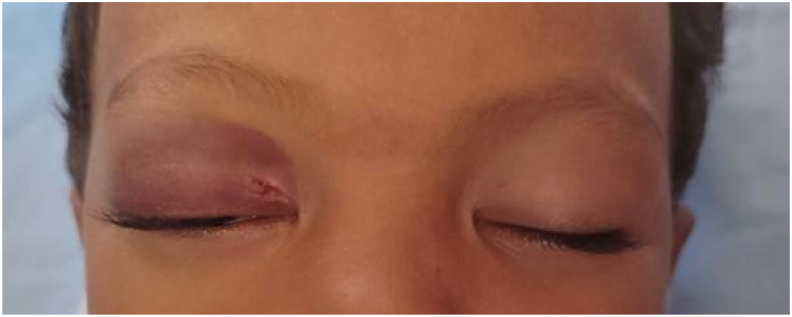
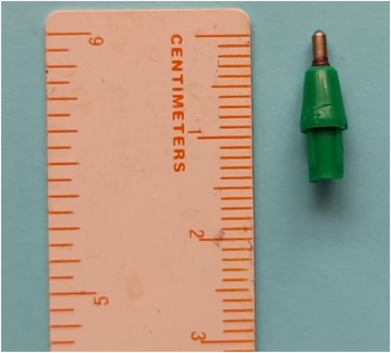
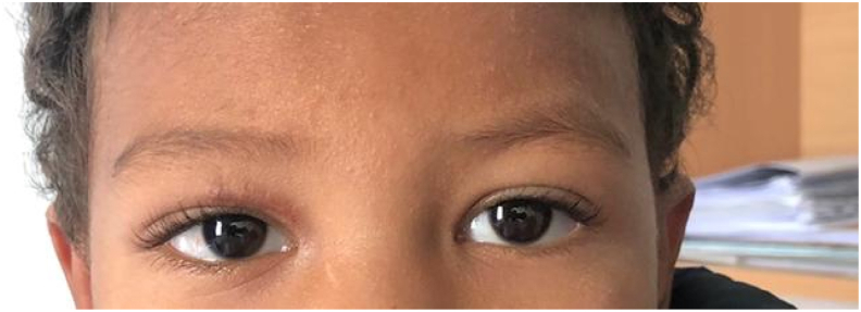
Apart from congenital causes, orbital trauma is a leading cause of unilateral vision loss in children. We report the case of a 2-year-old child who was victim of an orbital trauma of the right eye caused by a ballpoint pen. He consulted us the day after the trauma with significant palpebral edema making the examination difficult. An emergency CT scan of the orbit and brain showed the presence of a right intraorbital foreign body. The patient underwent removal of the foreign body by an anterior orbitotomy with general antibiotic therapy and a simple postoperative course. Penetrating trauma to the orbit should raise the suspicion of the presence of a foreign body. A CT scan should be performed to specify its location. The extraction of the foreign body can be a challenge that requires an experienced surgical team.
Keywords: Anterior orbitotomy; CT scan; Case report; Child; Intraorbital foreign body.
© 2022 The Authors.
Conflict of interest statement
No conflict of interest exists. We wish to confirm that there are no known conflicts of interest associated with this publication and there has been no significant financial support for this work that could have influenced its outcome.
Figures


References
-
- Agha R.A., Franchi T., Sohrabi C., Mathew G., for the SCARE Group The SCARE 2020 guideline: updating consensus surgical CAse REport (SCARE) guidelines. Int. J. Surg. 2020;84:226–230. - PubMed
Publication types
LinkOut - more resources
Full Text Sources
