Predicting Optical Coherence Tomography-Derived High Myopia Grades From Fundus Photographs Using Deep Learning
- PMID: 35308524
- PMCID: PMC8927672
- DOI: 10.3389/fmed.2022.842680
Predicting Optical Coherence Tomography-Derived High Myopia Grades From Fundus Photographs Using Deep Learning
Abstract
Purpose: To develop an artificial intelligence (AI) system that can predict optical coherence tomography (OCT)-derived high myopia grades based on fundus photographs.
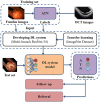
Methods: In this retrospective study, 1,853 qualified fundus photographs obtained from the Zhongshan Ophthalmic Center (ZOC) were selected to develop an AI system. Three retinal specialists assessed corresponding OCT images to label the fundus photographs. We developed a novel deep learning model to detect and predict myopic maculopathy according to the atrophy (A), traction (T), and neovascularisation (N) classification and grading system. Furthermore, we compared the performance of our model with that of ophthalmologists.
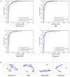
Results: When evaluated on the test set, the deep learning model showed an area under the receiver operating characteristic curve (AUC) of 0.969 for category A, 0.895 for category T, and 0.936 for category N. The average accuracy of each category was 92.38% (A), 85.34% (T), and 94.21% (N). Moreover, the performance of our AI system was superior to that of attending ophthalmologists and comparable to that of retinal specialists.
Conclusion: Our AI system achieved performance comparable to that of retinal specialists in predicting vision-threatening conditions in high myopia via simple fundus photographs instead of fundus and OCT images. The application of this system can save the cost of patients' follow-up, and is more suitable for applications in less developed areas that only have fundus photography.
Keywords: artificial intelligence; deep learning; fundus photographs; high myopia; optical coherence tomography.
Copyright © 2022 Wu, Cai, Xie, Chen, Wang, Lei, Zheng and Lu.
Conflict of interest statement
The authors declare that the research was conducted in the absence of any commercial or financial relationships that could be construed as a potential conflict of interest.
Figures


Similar articles
-
Development and validation of a deep learning system to screen vision-threatening conditions in high myopia using optical coherence tomography images.Br J Ophthalmol. 2022 May;106(5):633-639. doi: 10.1136/bjophthalmol-2020-317825. Epub 2020 Dec 21. Br J Ophthalmol. 2022. PMID: 33355150 Free PMC article.
-
Automatic Screening and Identifying Myopic Maculopathy on Optical Coherence Tomography Images Using Deep Learning.Transl Vis Sci Technol. 2021 Nov 1;10(13):10. doi: 10.1167/tvst.10.13.10. Transl Vis Sci Technol. 2021. PMID: 34751744 Free PMC article.
-
Enhancing pathological myopia diagnosis: a bimodal artificial intelligence approach integrating fundus and optical coherence tomography imaging for precise atrophy, traction and neovascularisation grading.Br J Ophthalmol. 2025 May 20:bjo-2024-326252. doi: 10.1136/bjo-2024-326252. Online ahead of print. Br J Ophthalmol. 2025. PMID: 40393796
-
Artificial Intelligence in Myopic Maculopathy: A Comprehensive Review of Identification, Classification, and Monitoring Using Diverse Imaging Modalities.Cureus. 2025 Feb 7;17(2):e78685. doi: 10.7759/cureus.78685. eCollection 2025 Feb. Cureus. 2025. PMID: 40062093 Free PMC article. Review.
-
Artificial Intelligence in Predicting Systemic Parameters and Diseases From Ophthalmic Imaging.Front Digit Health. 2022 May 26;4:889445. doi: 10.3389/fdgth.2022.889445. eCollection 2022. Front Digit Health. 2022. PMID: 35706971 Free PMC article. Review.
Cited by
-
Predicting visual acuity of treated ocular trauma based on pattern visual evoked potentials by machine learning models.Front Cell Dev Biol. 2025 Aug 1;13:1619956. doi: 10.3389/fcell.2025.1619956. eCollection 2025. Front Cell Dev Biol. 2025. PMID: 40823531 Free PMC article.
-
Global trends and hotspots in artificial intelligence for high myopia: a bibliometric analysis.Front Med (Lausanne). 2025 May 9;12:1567440. doi: 10.3389/fmed.2025.1567440. eCollection 2025. Front Med (Lausanne). 2025. PMID: 40417679 Free PMC article.
-
Artificial intelligence-aided diagnosis and treatment in the field of optometry.Int J Ophthalmol. 2023 Sep 18;16(9):1406-1416. doi: 10.18240/ijo.2023.09.06. eCollection 2023. Int J Ophthalmol. 2023. PMID: 37724269 Free PMC article.
-
A Beginner's Guide to Artificial Intelligence for Ophthalmologists.Ophthalmol Ther. 2024 Jul;13(7):1841-1855. doi: 10.1007/s40123-024-00958-3. Epub 2024 May 11. Ophthalmol Ther. 2024. PMID: 38734807 Free PMC article. Review.
-
Artificial intelligence in pathologic myopia: a review of clinical research studies.Front Med (Lausanne). 2025 Apr 23;12:1572750. doi: 10.3389/fmed.2025.1572750. eCollection 2025. Front Med (Lausanne). 2025. PMID: 40337273 Free PMC article.
References
-
- Fricke TR, Jong M, Naidoo KS, Sankaridurg P, Naduvilath TJ, Ho SM, et al. . Global prevalence of visual impairment associated with myopic macular degeneration and temporal trends from 2000 through 2050: systematic review, meta-analysis and modelling. Br J Ophthalmol. (2018) 102:855–62. 10.1136/bjophthalmol-2017-311266 - DOI - PMC - PubMed
LinkOut - more resources
Full Text Sources

