Review
. 2022 Mar 3:16:828532.
doi: 10.3389/fnsys.2022.828532. eCollection 2022.

How Does the Central Nervous System for Posture and Locomotion Cope With Damage-Induced Neural Asymmetry?

Affiliations
Review

How Does the Central Nervous System for Posture and Locomotion Cope With Damage-Induced Neural Asymmetry?

Didier Le Ray et al. Front Syst Neurosci. .

Abstract

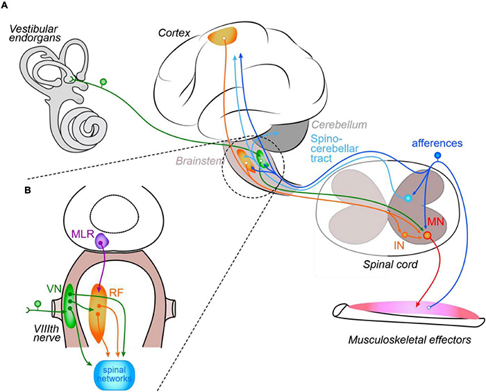
In most vertebrates, posture and locomotion are achieved by a biomechanical apparatus whose effectors are symmetrically positioned around the main body axis. Logically, motor commands to these effectors are intrinsically adapted to such anatomical symmetry, and the underlying sensory-motor neural networks are correspondingly arranged during central nervous system (CNS) development. However, many developmental and/or life accidents may alter such neural organization and acutely generate asymmetries in motor operation that are often at least partially compensated for over time. First, we briefly present the basic sensory-motor organization of posturo-locomotor networks in vertebrates. Next, we review some aspects of neural plasticity that is implemented in response to unilateral central injury or asymmetrical sensory deprivation in order to substantially restore symmetry in the control of posturo-locomotor functions. Data are finally discussed in the context of CNS structure-function relationship.

Keywords: development; injury; motor recovery; neuronal networks; sensory-motor integration.


Conflict of interest statement

The authors declare that the research was conducted in the absence of any commercial or financial relationships that could be construed as a potential conflict of interest.

Figures

FIGURE 1
FIGURE 1
Schematic organization of the control of posture and locomotion in vertebrates. Illustrated central and peripheral structures and connecting pathways (A) are arbitrarily limited to those discussed in the review. The inset (B) focuses on the main brainstem structures responsible for posturo-locomotor supraspinal commands. IN, interneuron; MN, motoneuron; MLR, mesencephalic locomotor region; VN, vestibular nuclei; RF, reticular formation; VIIIth nerve, vestibular nerve.
FIGURE 2
FIGURE 2
Unilateral stroke-evoked corticospinal plasticity. (A) In normal rats, corticospinal neurons from sensory-motor areas (dark blue projections) specifically connect either cervical or lumbar spinal MNs respectively involved in fore- and hindlimb fine motricity. (B) After a focal stroke in the cortical region hosting neurons projecting into cervical segments (red star), forelimb-related corticospinal neurons degenerate (light blue), and hindlimb-related neurons normally projecting only onto lumbar MNs sprout collaterals that make synapse onto cervical MNs deprived of their normal corticospinal inputs.
FIGURE 3
FIGURE 3
Spinal network reorganization after unilateral spinal cord injury. (A) The symmetrical organization of uncrossed (light blue) and crossed (dark blue) corticospinal and reticulospinal (green) projections (illustrated in upper inset) and local sensory inputs (cyan) onto segmental INs (pink) and MN (orange) in control animal (A1) exhibit adaptations below spinal cord hemisection (A2) ipsilesional sensory inputs invade IN and MN dendritic territories deprived from their ipsilateral descending projections, and contralateral descending axons sprout terminals into the ipsilesional hemicord. See text for details. Cx, cortex; Bs, brainstem; Sc, spinal cord; iCS and dCS, ipsilateral and decussating corticospinal tracts; RS, reticulospinal. (B) SCI-induced acute modifications in motor neuron (MN) and excitatory and inhibitory interneurons (eIN and iIN, respectively) intrinsic and synaptic properties leading to MN hyper-excitability. Sensory-evoked responses are enhanced due to increased expression of NMDA receptors (NMDA-Rs) in both MN and eIN, and increased expression of serotonin receptors (5-HT2B/C-Rs) in eIN. In contrast, inhibitory influences from iIN onto MN are reduced due to lower motoneuronal expression of GABA and glycine receptors (GABA-Rs and Gly-Rs, respectively). In addition, overexpression of sodium/calcium permeant channels (Na+/Ca2+ chan.) and lower expression of potassium channels (K+ chan.) intrinsically increase MN excitability. The relative expression of membrane channels and receptors is illustrated as follows: one item shows a decrease and three items an increase compared to a “normal expression” of two items.
FIGURE 4
FIGURE 4
Unilateral vestibular deprivation-induced adaptations in central vestibular nuclei and cerebellum. (A) Acute effects. A unilateral loss of vestibular afferents (vest.) triggers a rapid decrease in ipsilesional CVN neurons activity due to conjoint intrinsic and metabolic modifications together with increased inhibition from contralateral central vestibular nuclei (CVNs) and ipsilateral cerebellar nuclei. Although intrinsic properties of CVN neurons change bilaterally, net effect consists of a disequilibrium in reciprocal inhibition through commissural vestibular pathways (cINs) resulting in a stronger inhibition of ipsilesional CVNs. vis., propr., visual and proprioceptive afferents; NO, nitric oxide. Dotted lines: underactivated influences. (B) Compensated cerebello-vestibular network. Restoration of bilateral balance results from additional changes in intrinsic and synaptic properties of CVN neurons on both sides, together with ipsilesional GABAergic neurogenesis (yellow dots) and increased synaptic weight of both visual and proprioceptive inputs onto ipsilesional CVNs. Bilateral balance restoration also occurs in cerebellar nuclei due to a relative increase in NO on the contralesional side.
FIGURE 5
FIGURE 5
Functional organization of the spinal posturo-locomotor network in the intact and vestibulo-lesioned Xenopus. (A) Schemes illustrating the symmetrical organization of the most basic brainstem/spinal cord sensory-motor network responsible for the control of locomotion and posture in intact pre-metamorphosis tadpole (left) and post-metamorphosis juvenile (right) Xenopus (adapted from Beyeler et al., 2008). (B) Acute (left) and chronic (time interval equivalent to metamorphosis duration; right) effects of a right-side UVD in the juvenile Xenopus. Note that vestibular endorgans/Scarpa ganglion removal generates a net ipsilesional over-excitation of the spinal network, which results in a permanently impaired locomotor behavior (i.e., rolling swimming toward the lesion side; adapted from Beyeler et al., 2013). (C) Acute UVD (left) triggers spinal imbalance and rolling behavior in tadpoles. In contrast, after the animals had metamorphosed (right) normal swimming is restored although bilateral descending commands remain imbalanced. Recovery is allowed by a symmetrical activation of the postural axial network resulting from the construction, during metamorphosis, of an asymmetrical propriospinal posturo-locomotor network (adapted from Beyeler et al., 2013). Thin lines, normal activation; thick lines, hyper-activation; vest., vestibular.

References

    1. Ahuja C. S., Wilson J. R., Nori S., Kotter M. R. N., Druschel C., Curt A., et al. (2017). Traumatic spinal cord injury. Nat. Rev. Dis. Primer 3:17018. 10.1038/nrdp.2017.18 - DOI - PubMed
    1. Akay T., Murray A. J. (2021). Relative contribution of proprioceptive and vestibular sensory systems to locomotion: opportunities for discovery in the age of molecular science. Int. J. Mol. Sci. 22:1467. 10.3390/ijms22031467 - DOI - PMC - PubMed
    1. Alizadeh A., Dyck S. M., Karimi-Abdolrezaee S. (2019). Traumatic spinal cord injury: an overview of pathophysiology, models and acute injury mechanisms. Front. Neurol. 10:282. 10.3389/fneur.2019.00282 - DOI - PMC - PubMed
    1. Allard P., Tabin C. J. (2009). Achieving bilateral symmetry during vertebrate limb development. Semin. Cell Dev. Biol. 20 479–484. 10.1016/j.semcdb.2008.10.011 - DOI - PubMed
    1. Alvarez F. J., Bullinger K. L., Titus H. E., Nardelli P., Cope T. C. (2010). Permanent reorganization of Ia afferent synapses on motoneurons after peripheral nerve injuries: monosynaptic stretch reflexes after nerve injuries. Ann. N. Y. Acad. Sci. 1198 231–241. 10.1111/j.1749-6632.2010.05459.x - DOI - PMC - PubMed

LinkOut - more resources