

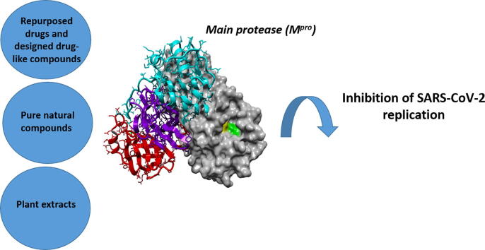
Inhibition of the main protease of SARS-CoV-2 (Mpro) by repurposing/designing drug-like substances and utilizing nature's toolbox of bioactive compounds
- PMID: 35308802
- PMCID: PMC8920478
- DOI: 10.1016/j.csbj.2022.03.009
Inhibition of the main protease of SARS-CoV-2 (Mpro) by repurposing/designing drug-like substances and utilizing nature's toolbox of bioactive compounds
Abstract
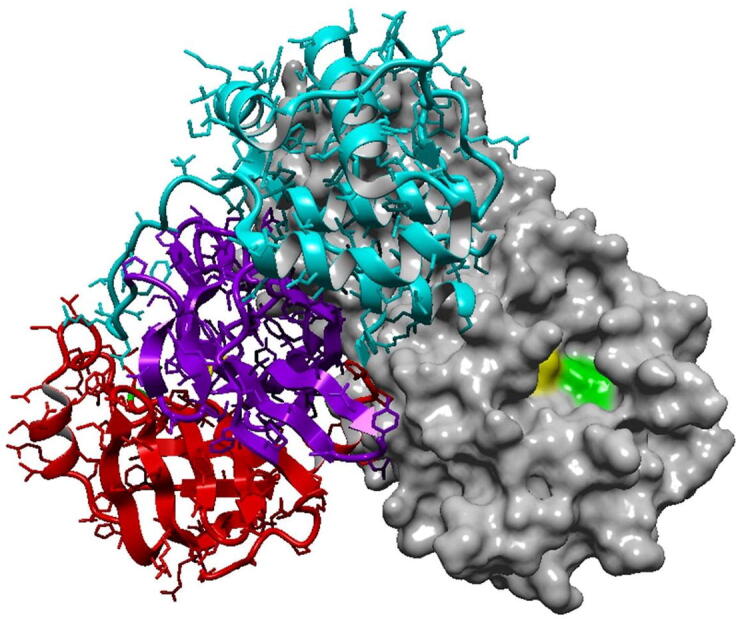
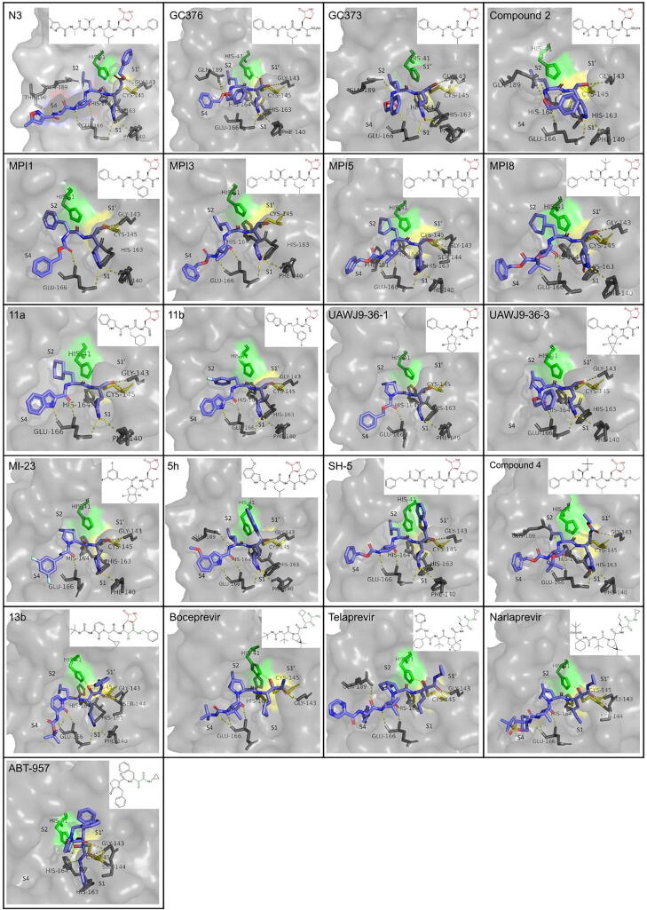
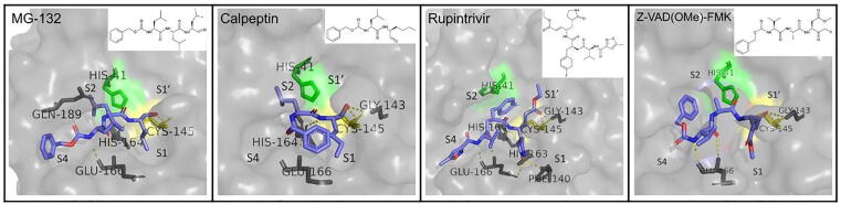
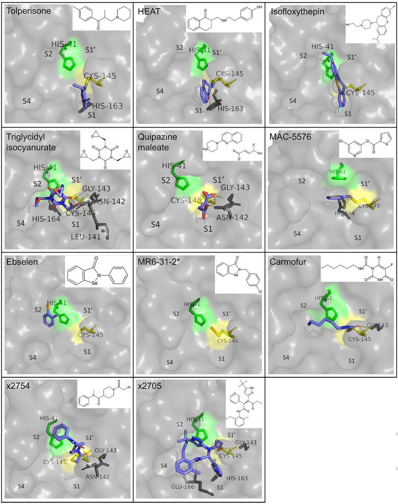
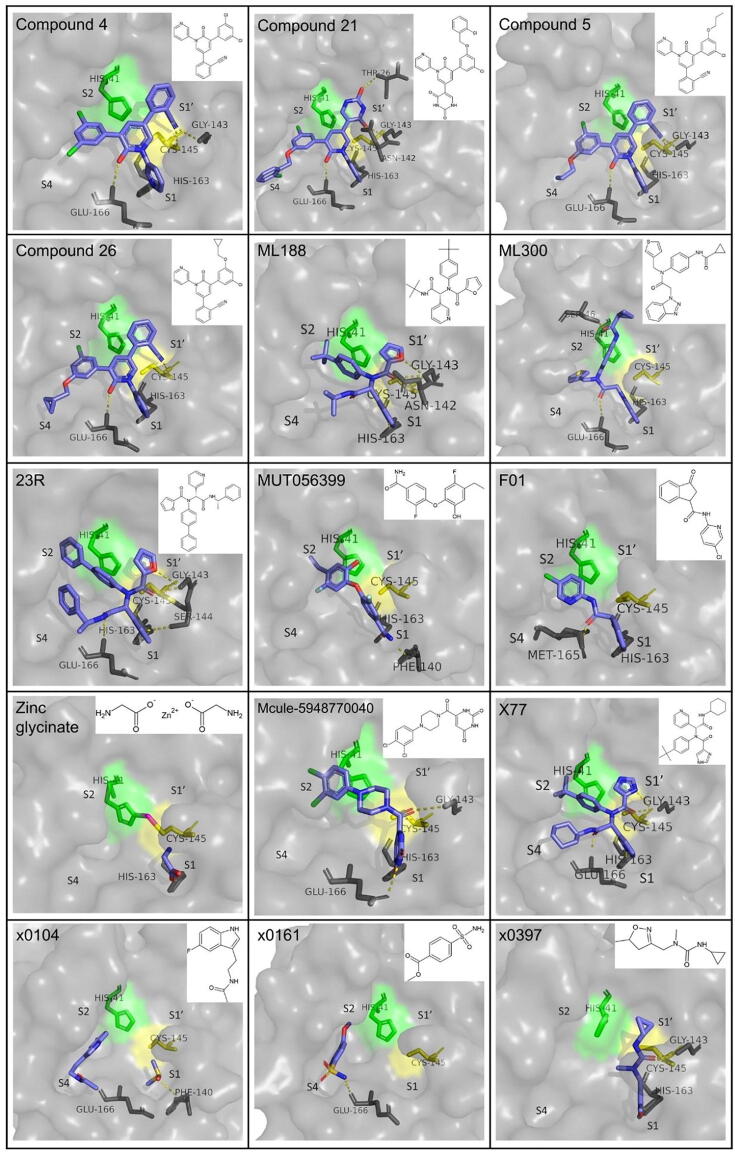
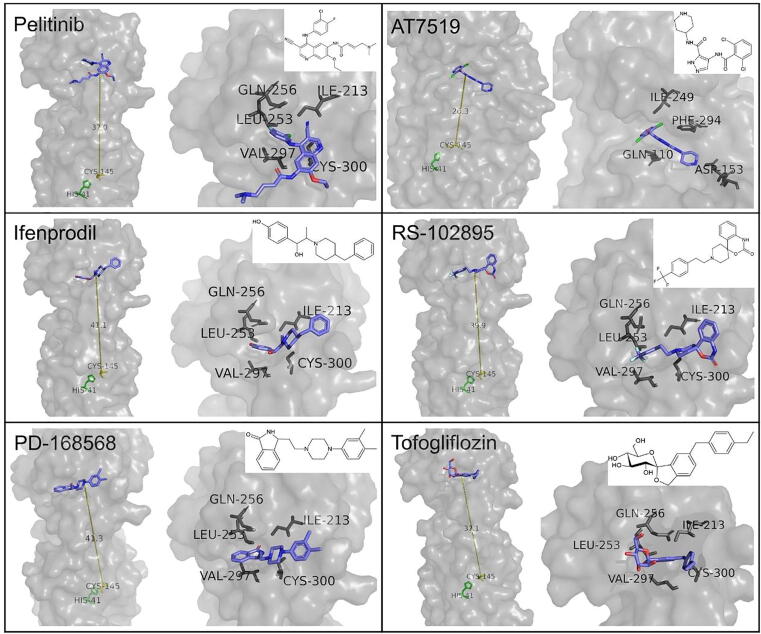
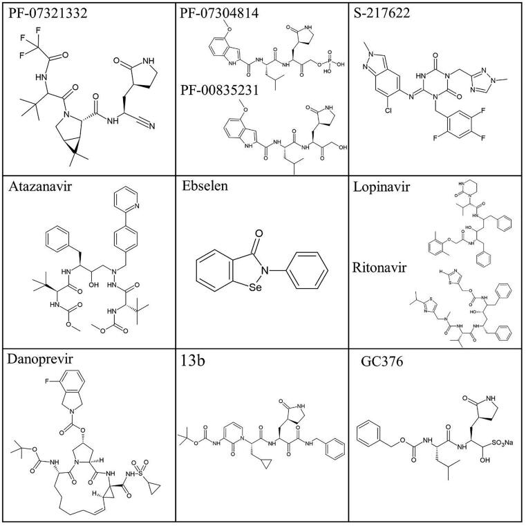
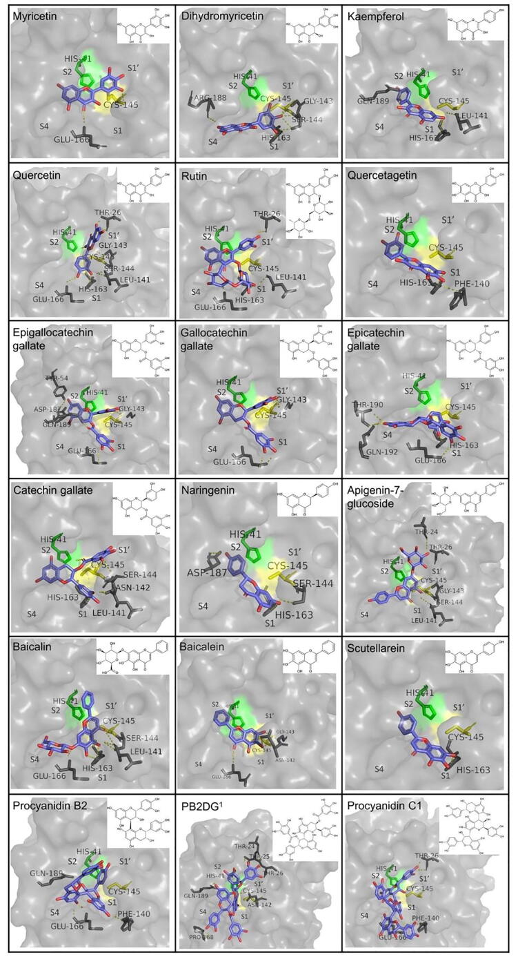
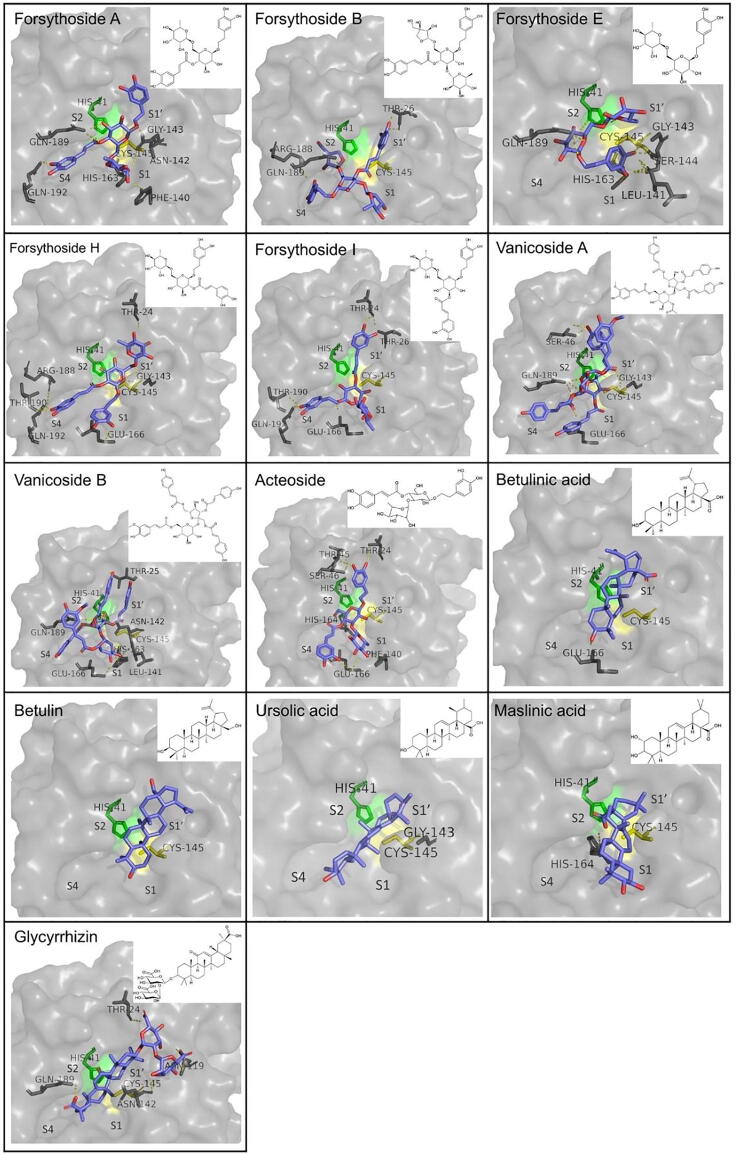
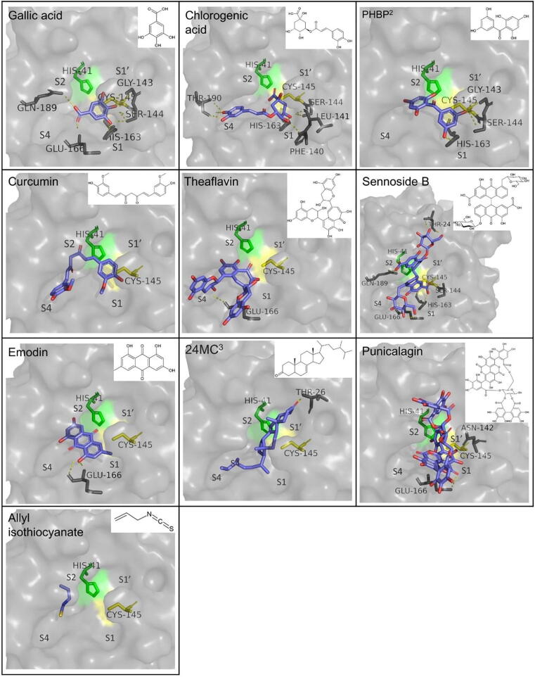
The emergence of the Severe Acute Respiratory Syndrome Coronavirus-2 (SARS-CoV-2) has resulted in a long pandemic, with numerous cases and victims worldwide and enormous consequences on social and economic life. Although vaccinations have proceeded and provide a valuable shield against the virus, the approved drugs are limited and it is crucial that further ways to combat infection are developed, that can also act against potential mutations. The main protease (Mpro) of the virus is an appealing target for the development of inhibitors, due to its importance in the viral life cycle and its high conservation among different coronaviruses. Several compounds have shown inhibitory potential against Mpro, both in silico and in vitro, with few of them also having entered clinical trials. These candidates include: known drugs that have been repurposed, molecules specifically designed based on the natural substrate of the protease or on structural moieties that have shown high binding affinity to the protease active site, as well as naturally derived compounds, either isolated or in plant extracts. The aim of this work is to collectively present the results of research regarding Mpro inhibitors to date, focusing on the function of the compounds founded by in silico simulations and further explored by in vitro and in vivo assays. Creating an extended portfolio of promising compounds that may block viral replication by inhibiting Mpro and by understanding involved structure-activity relationships, could provide a basis for the development of effective solutions against SARS-CoV-2 and future related outbreaks.
Keywords: Coronavirus; Enzyme inhibition; Extracts; Main Protease; Natural compounds; Repurposed drugs; SARS-CoV-2.
© 2022 Published by Elsevier B.V. on behalf of Research Network of Computational and Structural Biotechnology.
Conflict of interest statement
The authors declare that they have no known competing financial interests or personal relationships that could have appeared to influence the work reported in this paper.
Figures
References
-
- U.S. Food and Drug administration, “Know Your Treatment Options for COVID-19,” 2021. https://www.fda.gov/consumers/consumer-updates/know-your-treatment-optio....
Publication types
LinkOut - more resources
Full Text Sources
Other Literature Sources
Miscellaneous