

Three Classes of Antioxidant Defense Systems and the Development of Postmenopausal Osteoporosis
- PMID: 35309045
- PMCID: PMC8927967
- DOI: 10.3389/fphys.2022.840293
Three Classes of Antioxidant Defense Systems and the Development of Postmenopausal Osteoporosis
Abstract
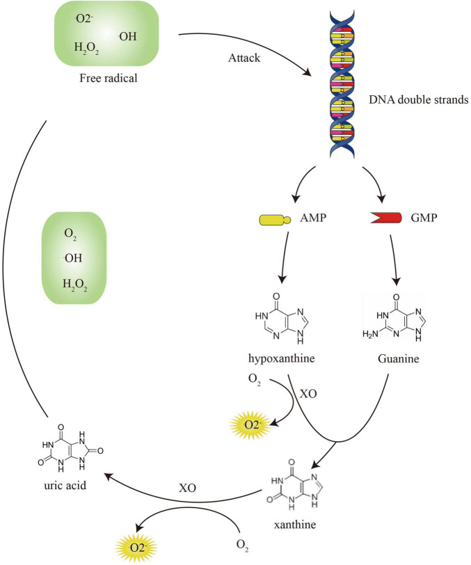
Osteoporosis is a common bone imbalance disease that threatens the health of postmenopausal women. Estrogen deficiency accelerates the aging of women. Oxidative stress damage is regarded as the main pathogenesis of postmenopausal osteoporosis. The accumulation of reactive oxygen species in the bone microenvironment plays a role in osteoblast and osteoclast apoptosis. Improving the oxidative state is essential for the prevention and treatment of postmenopausal osteoporosis. There are three classes of antioxidant defense systems in the body to eliminate free radicals and peroxides including antioxidant substances, antioxidant enzymes, and repair enzymes. In our review, we demonstrated the mechanism of antioxidants and their effect on bone metabolism in detail. We concluded that glutathione/oxidized glutathione (GSH/GSSG) conversion involved the PI3K/Akt-Nrf2/HO-1 signaling pathway and that the antioxidant enzyme-mediated mitochondrial apoptosis pathway of osteoblasts was necessary for the development of postmenopausal osteoporosis. Since the current therapeutic effects of targeting bone cells are not significant, improving the systemic peroxidation state and then regulating bone homeostasis will be a new method for the treatment of postmenopausal osteoporosis.
Keywords: GSH/GSSG; PI3K/AKT/Nrf2/HO-1; antioxidant system; oxidative stress; postmenopausal osteoporosis.
Copyright © 2022 Yang, Cao, Xue, Tao and Zhu.
Conflict of interest statement
The authors declare that the research was conducted in the absence of any commercial or financial relationships that could be construed as a potential conflict of interest.
Figures


References
-
- Alcendor R. R., Gao S., Zhai P., Zablocki D., Holle E., Yu X., et al. (2007). Sadoshima, Sirt1 regulates aging and resistance to oxidative stress in the heart. Circ. Res. 100 1512–1521. - PubMed
Publication types
LinkOut - more resources
Full Text Sources