

Updates in the Diagnosis of Intraductal Neoplasms of the Pancreas
- PMID: 35309060
- PMCID: PMC8931033
- DOI: 10.3389/fphys.2022.856803
Updates in the Diagnosis of Intraductal Neoplasms of the Pancreas
Erratum in
-
Corrigendum: Updates in the Diagnosis of Intraductal Neoplasms of the Pancreas.Front Physiol. 2022 May 13;13:923917. doi: 10.3389/fphys.2022.923917. eCollection 2022. Front Physiol. 2022. PMID: 35634156 Free PMC article.
Abstract
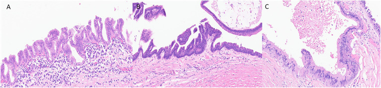
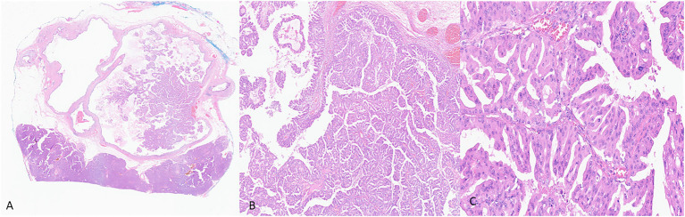
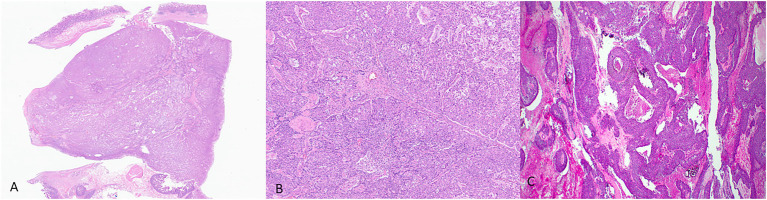
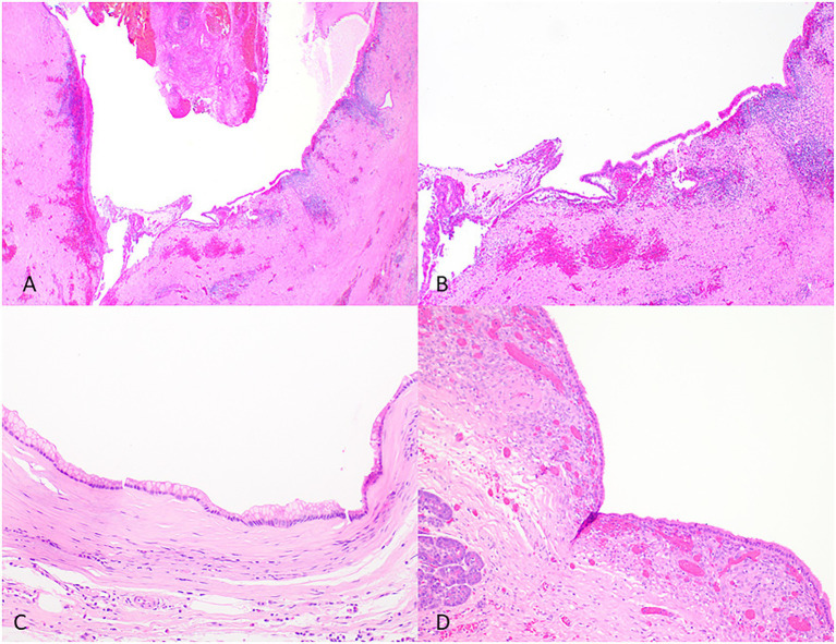
Pancreatic ductal adenocarcinoma (PDAC) is one of the deadliest types of cancer worldwide. There are many reasons for this dismal prognosis, including the advanced stage at the time of diagnosis and the lack of effective therapeutic approaches. Intraductal papillary mucinous neoplasms (IPMNs) represent detectable and treatable precursor lesions of PDAC. Our understanding of the pathology of IPMNs has evolved over the past few decades, and new advances in diagnostic tools have emerged. The new World Health Organization (WHO) classification scheme now recognizes the previously considered variants of IPMNs, such as intraductal oncocytic papillary neoplasms (IOPNs) and intraductal tubulopapillary neoplasms (ITPNs), as distinct neoplasms. New imaging and molecular diagnostic tests are being developed to recognize these PDAC precursor lesions better. Here, we review the advances in diagnostic tools for IPMNs, IOPNs, and ITPNs, emphasizing the new (5th edition, 2019) WHO classification for pathological diagnosis, molecular markers, new laboratory tests, and imaging tools.
Keywords: classification; intraductal oncocytic papillary neoplasm; intraductal papillary mucinous neoplasm; intraductal tubulopapillary neoplasm; pancreatic ductal adenocarcinoma.
Copyright © 2022 Assarzadegan, Babaniamansour and Shi.
Conflict of interest statement
The authors declare that the research was conducted in the absence of any commercial or financial relationships that could be construed as a potential conflict of interest.
Figures
References
-
- Adsay N. V., Merati K., Andea A., Sarkar F., Hruban R. H., Wilentz R. E., et al. (2002). The dichotomy in the preinvasive neoplasia to invasive carcinoma sequence in the pancreas: differential expression of MUC1 and MUC2 supports the existence of two separate pathways of carcinogenesis. Mod. Pathol. 15, 1087–1095. doi: 10.1097/01.MP.0000028647.98725.8B, PMID: - DOI - PubMed
-
- Adsay N. V., Merati K., Basturk O., Iacobuzio-Donahue C., Levi E., Cheng J. D., et al. (2004). Pathologically and biologically distinct types of epithelium in intraductal papillary mucinous neoplasms: delineation of an "intestinal" pathway of carcinogenesis in the pancreas. Am. J. Surg. Pathol. 28, 839–848. doi: 10.1097/00000478-200407000-00001, PMID: - DOI - PubMed
-
- Adsay V., Mino-Kenudson M., Furukawa T., Basturk O., Zamboni G., Marchegiani G., et al. (2016). Pathologic evaluation and reporting of intraductal papillary mucinous neoplasms of the pancreas and other tumoral intraepithelial neoplasms of pancreatobiliary tract: recommendations of Verona Consensus Meeting. Ann. Surg. 263, 162–177. doi: 10.1097/SLA.0000000000001173, PMID: - DOI - PMC - PubMed
Publication types
Grants and funding
LinkOut - more resources
Full Text Sources
Miscellaneous