. 2022 Mar 3:13:834988.
doi: 10.3389/fimmu.2022.834988. eCollection 2022.

Signaling Through FcγRIIA and the C5a-C5aR Pathway Mediate Platelet Hyperactivation in COVID-19

Collaborators, Affiliations

Signaling Through FcγRIIA and the C5a-C5aR Pathway Mediate Platelet Hyperactivation in COVID-19

Sokratis A Apostolidis et al. Front Immunol. .

Abstract

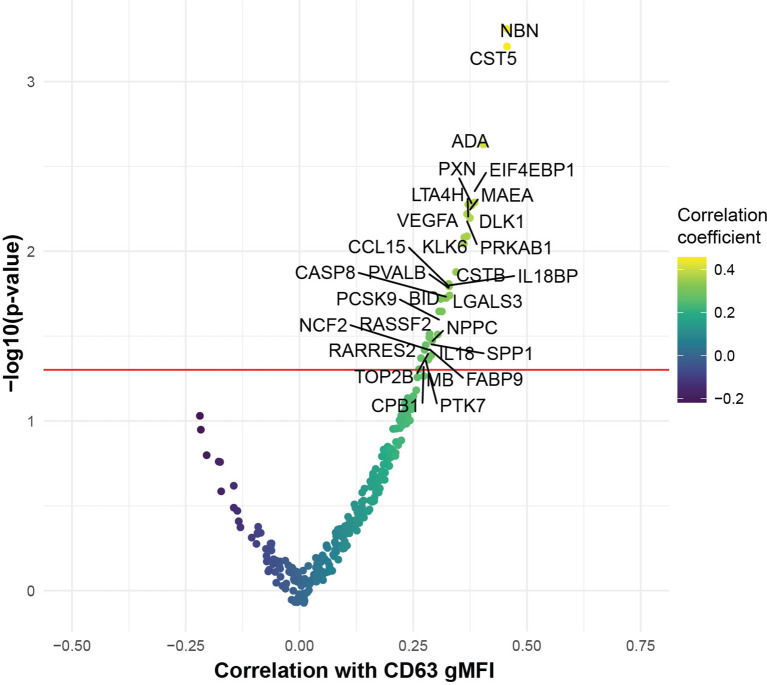
Patients with COVID-19 present with a wide variety of clinical manifestations. Thromboembolic events constitute a significant cause of morbidity and mortality in patients infected with SARS-CoV-2. Severe COVID-19 has been associated with hyperinflammation and pre-existing cardiovascular disease. Platelets are important mediators and sensors of inflammation and are directly affected by cardiovascular stressors. In this report, we found that platelets from severely ill, hospitalized COVID-19 patients exhibited higher basal levels of activation measured by P-selectin surface expression and had poor functional reserve upon in vitro stimulation. To investigate this question in more detail, we developed an assay to assess the capacity of plasma from COVID-19 patients to activate platelets from healthy donors. Platelet activation was a common feature of plasma from COVID-19 patients and correlated with key measures of clinical outcome including kidney and liver injury, and APACHEIII scores. Further, we identified ferritin as a pivotal clinical marker associated with platelet hyperactivation. The COVID-19 plasma-mediated effect on control platelets was highest for patients that subsequently developed inpatient thrombotic events. Proteomic analysis of plasma from COVID-19 patients identified key mediators of inflammation and cardiovascular disease that positively correlated with in vitro platelet activation. Mechanistically, blocking the signaling of the FcγRIIa-Syk and C5a-C5aR pathways on platelets, using antibody-mediated neutralization, IgG depletion or the Syk inhibitor fostamatinib, reversed this hyperactivity driven by COVID-19 plasma and prevented platelet aggregation in endothelial microfluidic chamber conditions. These data identified these potentially actionable pathways as central for platelet activation and/or vascular complications and clinical outcomes in COVID-19 patients. In conclusion, we reveal a key role of platelet-mediated immunothrombosis in COVID-19 and identify distinct, clinically relevant, targetable signaling pathways that mediate this effect.

Keywords: COVID - 19; FcγRIIa; complement; fostamatinib; platelet.


Conflict of interest statement

SH has received consultancy fees from Sanofi Pasteur, Lumen, Novavax, and Merck for work unrelated to this report. AH is a consultant for Immunai. EW is consulting or is an advisor for Merck, Elstar, Janssen, Related Sciences, Synthekine and Surface Oncology. EW is a founder of Surface Oncology and Arsenal Biosciences. EW is an inventor on a patent (US Patent number 10,370,446) submitted by Emory University that covers the use of PD-1 blockade to treat infections and cancer. NM reports funding to her institution from Quantum Leap Healthcare Collaborative, Athersys, Inc., Biomarck, Inc. The remaining authors declare that the research was conducted in the absence of any commercial or financial relationships that could be construed as a potential conflict of interest.

Figures

Graphical Abstract
Graphical Abstract
Figure 1
Figure 1
Platelets from hospitalized COVID-19 patients exhibit increased activation at baseline and diminished functional reserve. (A, B) Representative histograms (A) and cumulative data (B) for CD62P surface expression of ex vivo isolated platelets assayed with or without TRAP stimulation for 20mins. The CD62P gMFI ratio of TRAP/basal-treated is also shown. The three patient cohorts shown are COVID-19 inpatient (n=38), COVID-19 convalescent (n=23) and healthy donors (n=7). Kruskal-Wallis non-parametric testing was used to compare all three groups and pair-wise comparisons were also performed; p-values are depicted. (C) Cumulative data for CD62P surface expression of ex vivo isolated platelets at baseline (basal), after TRAP activation (TRAP) and their ratio for hospitalized COVID-19 patients on full-dose (n=10) or prophylactic/intermediate dose (n=13) of anticoagulation. Wilcoxon non-parametric testing was used and the p-values are depicted. (D) Cumulative data for CD62P surface expression of ex vivo isolated platelets at baseline for hospitalized COVID-19 patients with (n=31) or without (n=7) cardiovascular disease risk factors. Wilcoxon non-parametric testing was used, and the p-value is depicted. FMO control, Fluorescence minus one control; gMFI, geometric mean fluorescence intensity; TRAP, Thrombin Receptor Activation Peptide.
Figure 2
Figure 2
The ability of COVID19 plasma to activate platelets is increased for patients with evidence of organ damage, high circulating levels of ferritin, and at timepoints preceding a thrombotic event. (A) Plasma from COVID-19 inpatient (n=63), COVID-19 convalescent (n=20) and healthy donors (n=9) was incubated with platelets isolated from healthy volunteers. The gMFI levels of CD63, CD62P, CD32 and C3aR are shown in these three groups. Kruskal-Wallis non-parametric testing was used to compare all three groups and pair-wise comparisons were also performed; p-values are depicted. (B) Spearman correlation of the gMFI surface levels of the markers used in (A) and selected clinical parameters of the COVID-19 inpatient group (n=63). Δ correlation coefficient shown on the key (bottom). Asterisks *, ** and *** denote p values less than 0.05, 0.01 and 0.001 respectively. Highlighted squares denote FDR values less than 0.05. (C) CD63 gMFI surface levels of control platelets induced by plasma derived from COVID-19 patients with thrombosis drawn at different timepoints relative to the thrombotic event. (D) CD63 gMFI surface levels of control platelets induced by plasma derived from COVID-19 patients prior to thrombosis and after thrombosis, COVID-19 patients without thrombosis and COVID-19 patients in convalescence. Kruskal-Wallis non-parametric testing was used to compare all four groups and pair-wise comparisons were also performed using Wilcoxon testing; p-values are depicted. (E) Representative scatter plots for creatinine vs CD63, platelet count vs CD62P and APACHE III score vs CD63. Samples are colored based on the absence (grey color) or presence of thrombosis relative to the time of draw (red before thrombosis and blue after thrombosis). The Spearman correlation co-efficient and the corresponding p-value are depicted.
Figure 3
Figure 3
COVID-19 plasma induced platelet activation is associated with markers of inflammation and cardiovascular disease. Volcano plot with the x-axis representing the correlation coefficient of the different analytes with CD63 gMFI induction on platelets by COVID-19 plasma (n=53) and the y-axis depicting the -log10 transformation of the corresponding p-value.
Figure 4
Figure 4
FcγRIIa activation and complement anaphylatoxins mediate platelet activation in COVID-19. (A) Heatmap of gMFI expression of CD62P, CD63, CD32 and C3aR on the surface of control platelets incubated with COVID-19 plasma (n=10 patients) in the presence or absence of neutralizing antibodies to FcγRIIa, IL6, C3a and C5a, as indicated. (B) Boxplots of gMFI expression of CD63 on the surface of control platelets incubated with COVID-19 plasma (n=5 patients, depicted with distinct colors) with different combinations of neutralizing antibodies to FcγRIIa, IL6, C3a and C5a, as indicated. (C) Boxplots of gMFI expression of CD63 on the surface of control platelets incubated with COVID-19 plasma (n = 9 patients, depicted with distinct colors) with different combinations of IgG depletion and neutralizing antibodies to C3a and C5a.
Figure 5
Figure 5
Fostamatinib ameliorates the heightened activation of platelets induced by COVID-19 plasma. (A) Phospho-Syk and total Syk gMFI were measured with flow cytometry of control platelets incubated with COVID-19 plasma (n=3 patients) for 1 and 5 minutes. Incubation with healthy control plasma for 5 mins was used to normalize the data. (B) Representative studies of a hematoporphyrin-induced photochemical injury model in an endothelial-lined microfluidic channel. Images show platelet adhesion (in white) immediately after infusion of isolated washed healthy donor platelets, and 5 min, 10 min and 15 min after infusion. Shown is a representative study where plasma from a severe COVID-19 patient was added to the platelets. In the samples indicated, the Syk inhibitor R406, FcR blocking antibody, or an anti-C5a antibody were added prior to infusion. Direction of blood flow is indicated by arrows. Size bar indicates 100µ. (C) Overall data analysis from studies with plasma derived from 7 patients with severe COVID-19. The Y-axis shows accumulated platelets in each microfluidic lane done immediately after the isolated platelet suspension was added and after 5, 10 and 15 minutes (log10 scale, fold change over healthy control plasma flown in uninjured channels). (D) All 7 sets of experiments from D are shown at the 10-minute timepoint.

Update of

References

    1. Yang J, Chen X, Deng X, Chen Z, Gong H, Yan H, et al. . Disease Burden and Clinical Severity of the First Pandemic Wave of COVID-19 in Wuhan, China. Nat Commun (2020) 11. doi: 10.1038/s41467-020-19238-2 - DOI - PMC - PubMed
    1. Zhou F, Yu T, Du R, Fan G, Liu Y, Liu Z, et al. . Clinical Course and Risk Factors for Mortality of Adult Inpatients With COVID-19 in Wuhan, China: A Retrospective Cohort Study. Lancet (2020) 395:1054–62. doi: 10.1016/S0140-6736(20)30566-3 - DOI - PMC - PubMed
    1. Huang C, Wang Y, Li X, Ren L, Zhao J, Hu Y, et al. . Clinical Features of Patients Infected With 2019 Novel Coronavirus in Wuhan, China. Lancet (2020) 395:497–506. doi: 10.1016/S0140-6736(20)30183-5 - DOI - PMC - PubMed
    1. Stadlbauer D, Amanat F, Chromikova V, Jiang K, Strohmeier S, Arunkumar GA, et al. . SARS-CoV-2 Seroconversion in Humans: A Detailed Protocol for a Serological Assay, Antigen Production, and Test Setup. Curr Protoc Microbiol (2020) 57:e100. doi: 10.1002/cpmc.100 - DOI - PMC - PubMed
    1. Middeldorp S, Coppens M, van Haaps TF, Foppen M, Vlaar AP, Müller MCA, et al. . Incidence of Venous Thromboembolism in Hospitalized Patients With COVID-19. J Thromb Haemost (2020) 18:1995–2002. doi: 10.1111/jth.14888 - DOI - PMC - PubMed

Publication types

MeSH terms