. 2022 Mar 2:10:830432.
doi: 10.3389/fcell.2022.830432. eCollection 2022.

Modeling Human Primary Microcephaly With hiPSC-Derived Brain Organoids Carrying CPAP-E1235V Disease-Associated Mutant Protein

Affiliations

Modeling Human Primary Microcephaly With hiPSC-Derived Brain Organoids Carrying CPAP-E1235V Disease-Associated Mutant Protein

Hsiao-Lung An et al. Front Cell Dev Biol. .

Abstract

The centrosome is composed of a pair of centrioles and serves as the major microtubule-organizing center (MTOC) in cells. Centrosome dysfunction has been linked to autosomal recessive primary microcephaly (MCPH), which is a rare human neurodevelopmental disorder characterized by small brain size with intellectual disability. Recently, several mouse models carrying mutated genes encoding centrosomal proteins have been generated to address the genotype-phenotype relationships in MCPH. However, several human-specific features were not observed in the mouse models during brain development. Herein, we generated isogenic hiPSCs carrying the gene encoding centrosomal CPAP-E1235V mutant protein using the CRISPR-Cas9 genome editing system, and examined the phenotypic features of wild-type and mutant hiPSCs and their derived brain organoids. Our results showed that the CPAP-E1235V mutant perturbed the recruitment of several centriolar proteins involved in centriole elongation, including CEP120, CEP295, CENTROBIN, POC5, and POC1B, onto nascent centrioles, resulting in the production of short centrioles but long cilia. Importantly, our wild-type hiPSC-derived brain organoid recapitulated many cellular events seen in the developing human brain, including neuronal differentiation and cortical spatial lamination. Interestingly, hiPSC-CPAP-E1235V-derived brain organoids induced p53-dependent neuronal cell death, resulting in the production of smaller brain organoids that mimic the microcephaly phenotype. Furthermore, we observed that the CPAP-E1235V mutation altered the spindle orientation of neuronal progenitor cells and induced premature neuronal differentiation. In summary, we have shown that the hiPSC-derived brain organoid coupled with CRISPR/Cas9 gene editing technology can recapitulate the centrosome/centriole-associated MCPH pathological features. Possible mechanisms for MCPH with centriole/centrosome dysfunction are discussed.

Keywords: CENPJ; CPAP; centriole duplication; cerebral organoid model; ciliopathy; human microcephaly; microcephaly gene mutations.


Conflict of interest statement

The authors declare that the research was conducted in the absence of any commercial or financial relationships that could be construed as a potential conflict of interest.

Figures
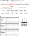
FIGURE 1
FIGURE 1
Generation of CRISPR/Cas9-mediated homozygous point mutation in the CPAP/CENPJ gene. (A) The scheme of homologous recombination between endogenous CPAP wild-type (WT) and exogenous repair template (single strain DNA, ssDNA) harbored A>T point mutation. Black inverted triangle: Cas9-cutting site. (B) Sanger sequencing results of two independent mutant clones displaying homozygous A>T point mutation in exon 16 of CPAP, which resulted in Glutamic acid (E) being replaced by Valine (V) at amino acid 1235. (C) CPAP protein expression level in WT and two independent mutant hiPSC clones. Black star symbol indicated non-specific bands.
FIGURE 2
FIGURE 2
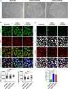
Characterization of CPAP-E1235V mutant hiPSC clones. (A) Bright field colony morphology of CPAP-WT and two mutant cell clones. (B) Immunofluorescent staining and quantification for pluripotency markers: NANOG (green), OCT4 (red), and DNA (DPAI, blue). n = 64 for CPAP-WT; n = 37 for CPAP-E1235V#1; n = 36 for CPAP-E1235V#2. Data represent mean ±SEM. (C) Staining and quantified results of apoptosis marker TUNEL in WT and mutant hiPSC clones. n = 664 for CPAP-WT; n = 281 for CPAP-E1235V#1; n = 619 for CPAP-E1235V#2. Data are presented as the mean ±SEM from a pool of n cells from three independent experiments. n.s.: not significant; **p < 0.01; ***p < 0.001. Scale bar: 200 μm in (A), 20 μm in (B,C).
FIGURE 3
FIGURE 3
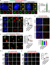
hiPSCs carrying CPAP-E1235V mutation displayed short centrioles and abnormally long cilia. Cells were synchronized at early S phase by treating aphidicolin (2 μg/ml) for 24 h (S phase) and released in fresh culture medium without aphidicolin for another 16 h (G2 phase). (A) Centriole numbers in control and two hiPSC mutant clones. Cells in the G2 phase were immunostained with antibodies against CENTRIN-2 (a centriole marker) and Acetylated-TUBULIN (Ac-TUB) (left) and the number of centriole was quantified (right) from three independent experiments. (B) Immunostaining and quantification of CPAP signals in S phase centrioles. n = 61 for CPAP-WT; n = 30 for CPAP-E1235V#1; n = 37 for CPAP-E1235V#2. (C) Airyscan images of centriole lengths by measuring the distance between two CEP162 dots in each pair of centrioles. n = 192 for CPAP-WT; n = 134 for CPAP-E1235V#1; n = 122 for CPAP-E1235V#2. (D,E) Immunostaining of CPAP-WT and mutant cells with antibodies against ARL13B (a ciliary membrane marker) and Ac-TUB (a centriole and ciliary axoneme marker). The cells were treated with 0.5% low serum starvation for 48 h. (D) The number of ciliated cells. n = 265 for CPAP-WT; n = 95 for CPAP-E1235V#1; n = 196 for CPAP-E1235V#2. Data are presented as the mean ±SEM from a pool of n cells from three independent experiments. (E) Quantification of ciliary length. n = 20 for CPAP-WT; n = 20 for CPAP-E1235V#1; n = 22 for CPAP-E1235V#2. Two types of long cilia were observed in the mutant cells. One type of long cilia displays long ciliary membranes (ARL13B+) and long ciliary axonemes (Ac-TUB+) (Ei), while the other type of cilia exhibit long ciliary membranes (ARL13B+) but short axonemes (Ac-TUB+) (Eii). Data are presented as the mean ±SEM. ***p < 0.001. Scale bar: 5 μm in (A,B,E), 0.5 μm in (C), 10 μm in (D).
FIGURE 4
FIGURE 4
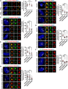
CPAP-E1235V mutation impaired the recruitment of centriole elongation proteins onto the centrioles. Cells were synchronized in the early S phase by treatment with aphidicolin (2 μg/ml) for 24 h. They were then fixed immediately with methanol (S phase centrioles) or released into fresh culture medium without aphidicolin for another 16 h (G2 phase centrioles). CPAP-WT or mutant cells in the S phase or G2 phase were immunostained with antibodies against the following centriolar proteins. (A) STIL, n = 42 for CPAP-WT; n = 25 for CPAP-E1235V#1; n = 34 for CPAP-E1235V#2. (B) CEP120, n = 50 for CPAP-WT; n = 42 for CPAP-E1235V#1; n = 48 for CPAP-E1235V#2. (C) CENTROBIN, n = 40 for all groups. (D) CEP295, n = 40 for all groups. (E) POC5, n = 42 for CPAP-WT; n = 40 for CPAP-E1235V#1; n = 40 for CPAP-E1235V#2. (F) POC1B, n = 40 for all groups. (G) CEP164, n = 88 for CPAP-WT; n = 74 for CPAP-E1235V#1; n = 38 for CPAP-E1235V#2. All data are presented as mean ±SEM from a pool of n cells from three independent experiments. n.s: not significant; **p < 0.01; ***p < 0.001. Scale bar: 5 μm.
FIGURE 5
FIGURE 5
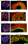
Characterization of hiPSC-derived brain organoid at Day 27 after culture. Immunostaining of the cryosections obtained from d27 organoids for (A) TUJ1 (a neuron marker)/PAX6 (a NPC marker), (B) Ki67 (a cell proliferating marker)/PAX6, (C) ZO-1 (a tight junction marker)/PAX6, and (D) ARL13B (a ciliary membrane marker)/CENTRIN-3 (a centriole marker). The cilia protruded from the apical surface in 27-day-old brain organoids. Scale bar: 200 μm in (A–D), 10 μm in enlarge (D) (right). White stars in (A) indicate the ventricle-like cavities in the d27 organoids. The enlarged image in [(A), right] is a 90-degree left turn from the left image.
FIGURE 6
FIGURE 6
hiPSCs-derived brain organoids recapitulate many cellular features of the developing human brain. (A) Neurogenesis in developing brain organoids. Immunostaining of the cryosection of developing organoids at the indicated time points for TUJ1 (a neuron marker)/PAX6 (a NPC marker). (B) Immunostaining of the 46-day-old brain organoid with antibodies against TBR1 (an early-born neural marker)/PAX6. (C,D) Immunostaining of developing organoids at the indicated time points for SATB2 (a later-born neural marker)/TBR1 (C) and for REELIN (a Cajal–Retzius cell marker)/TBR1 (D). White stars: lateral ventricle-like cavities. Scale bar: 50 μm in (A,C,D); 200 μm in (B).
FIGURE 7
FIGURE 7
CPAP-E1235V mutant organoids revealed the microcephaly phenotype. (A) Bright-filed morphology of CPAP-WT and mutant brain organoids at the embryoid body stage (Day 1 and Day 6) and differentiation stage (Day 27). The pool of larger brain organoids from WT and mutant groups was quantified and shown in right. n= 19 for CPAP-WT; n = 21 for CPAP-E1235V#1; n= 22 for CPAP-E1235V#2. Data are presented as mean ±SEM. (B) Immunostaining and quantitation of cleaved CASPASE 3 positive cells in 27d-old WT and mutant brain organoids. n = 5 for CPAP-WT; n = 4 for CPAP-E1235V#1; n = 4 for CPAP-E1235V#2. All data are presented as mean ±SEM from three independent experiments. (C) Immunostaining and quantification of p53 positive cells in 27-day-old WT and mutant brain organoids. n = 4 for CPAP-WT; n = 3 for CPAP-E1235V#1; n = 3 for CPAP-E1235V#2. All data are presented as mean ±SEM from three independent experiments. n. s: not significant; **p < 0.01; ***p < 0.001. Scale bar: 200 μm in (A), 20 μm in (B), 50 μm in (C).
FIGURE 8
FIGURE 8
CPAP-E1235V-derived brain organoid exhibited premature neuronal differentiation. (A) Immunostaining of TUJ1-positive neurons and PAX6-positive NPCs in 27-day-old WT and mutant brain organoids. The thickness of TUJ1-positive and PAX6-positive cell layers in 27-day-old WT and mutant organoids were quantified and are shown in (B). n = 8 for CPAP-WT; n = 6 for CPAP-E1235V#1; n = 5 for CPAP-E1235V#2. (C,D) Immunostaining of TUJ1-positive neurons and PAX6-positive NPCs (C) and their corresponding cell layer thickness in 52-day-old WT and mutant brain organoids were quantified and are shown in (D). n = 5 for CPAP-WT; n = 4 for CPAP-E1235V#1; n = 4 for CPAP-E1235V#2. All data are presented as mean ±SEM. *p < 0.05; **p < 0.01; ***p < 0.001. Scale bar: 50 μm in (A), 100 μm in (C).
FIGURE 9
FIGURE 9
CPAP-E1235V mutation induces NPC spindle misorientation in mutant brain organoids. (A) Representative images of NPC division in 27-day-old WT and mutant brain organoids with horizontal, oblique, and vertical mitotic spindle orientations at the ventricular surface (white lines). The spindle angle of mitotic anaphase NPCs (labeled by p-VIMENTIN) was calculated using a line (yellow) connecting two centrosomes (labelled by PERICENTRIN) and another line (white) representing the apical surface using the Image J software. Part of the PERICENTRIN-labelled centrosomes in NPCs is not visible due to the image being obtained from a single Z-stack (0.8 μm thickness), but not the merged Z-stacks. (B,C) Quantification of the spindle angle of the mitotic anaphase NPCs in 27-day-old CPAP-WT and mutant organoids. CPAP-WT NPCs divided horizontally in the majority of cases (0–30° angle), whereas mutant NPCs displayed many vertical orientations (C). Results were obtained from a pool of NPCs (n) from at least three independently collected organoids in each group. n = 21 NPCs for CPAP-WT; n = 19 NPCs for CPAP-E1235V#1; n = 19 NPCs for CPAP-E1235V#2 (C). Data are presented as mean ±SEM. ***p < 0.001. Scale bar: 10 μm.

Similar articles

Cited by

References

    1. Bhaduri A., Andrews M. G., Mancia Leon W., Jung D., Shin D., Allen D., et al. (2020). Cell Stress in Cortical Organoids Impairs Molecular Subtype Specification. Nature 578, 142–148. 10.1038/s41586-020-1962-0 - DOI - PMC - PubMed
    1. Bond J., Roberts E., Springell K., Lizarraga S., Scott S., Higgins J., et al. (2005). A Centrosomal Mechanism Involving CDK5RAP2 and CENPJ Controls Brain Size. Nat. Genet. 37, 353–355. 10.1038/ng1539 - DOI - PubMed
    1. Breslow D. K., Holland A. J. (2019). Mechanism and Regulation of Centriole and Cilium Biogenesis. Annu. Rev. Biochem. 88, 691–724. 10.1146/annurev-biochem-013118-111153 - DOI - PMC - PubMed
    1. Chang C. W., Hsu W. B., Tsai J. J., Tang C. J., Tang T. K. (2016). CEP295 Interacts with Microtubules and Is Required for Centriole Elongation. J. Cel Sci. 129, 2501–2513. 10.1242/jcs.186338 - DOI - PMC - PubMed
    1. Chen H.-Y., Wu C.-T., Tang C.-J. C., Lin Y.-N., Wang W.-J., Tang T. K. (2017). Human Microcephaly Protein RTTN Interacts with STIL and Is Required to Build Full-Length Centrioles. Nat. Commun. 8, 247. 10.1038/s41467-017-00305-0 - DOI - PMC - PubMed