. 2022 Mar 3:10:836797.
doi: 10.3389/fcell.2022.836797. eCollection 2022.

The Collagen Receptor Discoidin Domain Receptor 1b Enhances Integrin β1-Mediated Cell Migration by Interacting With Talin and Promoting Rac1 Activation

Affiliations

The Collagen Receptor Discoidin Domain Receptor 1b Enhances Integrin β1-Mediated Cell Migration by Interacting With Talin and Promoting Rac1 Activation

Corina M Borza et al. Front Cell Dev Biol. .

Abstract

Integrins and discoidin domain receptors (DDRs) 1 and 2 promote cell adhesion and migration on both fibrillar and non fibrillar collagens. Collagen I contains DDR and integrin selective binding motifs; however, the relative contribution of these two receptors in regulating cell migration is unclear. DDR1 has five isoforms (DDR1a-e), with most cells expressing the DDR1a and DDR1b isoforms. We show that human embryonic kidney 293 cells expressing DDR1b migrate more than DDR1a expressing cells on DDR selective substrata as well as on collagen I in vitro. In addition, DDR1b expressing cells show increased lung colonization after tail vein injection in nude mice. DDR1a and DDR1b differ from each other by an extra 37 amino acids in the DDR1b cytoplasmic domain. Interestingly, these 37 amino acids contain an NPxY motif which is a central control module within the cytoplasmic domain of β integrins and acts by binding scaffold proteins, including talin. Using purified recombinant DDR1 cytoplasmic tail proteins, we show that DDR1b directly binds talin with higher affinity than DDR1a. In cells, DDR1b, but not DDR1a, colocalizes with talin and integrin β1 to focal adhesions and enhances integrin β1-mediated cell migration. Moreover, we show that DDR1b promotes cell migration by enhancing Rac1 activation. Mechanistically DDR1b interacts with the GTPase-activating protein (GAP) Breakpoint cluster region protein (BCR) thus reducing its GAP activity and enhancing Rac activation. Our study identifies DDR1b as a major driver of cell migration and talin and BCR as key players in the interplay between integrins and DDR1b in regulating cell migration.

Keywords: Rac1; extracellular matrix; integrins; migration; receptor activation; receptor tyrosine kinase.


Conflict of interest statement

The authors declare that the research was conducted in the absence of any commercial or financial relationships that could be construed as a potential conflict of interest.

Figures

FIGURE 1
FIGURE 1
DDR1b supports lung colonization in vivo. (A) HEK cells were transfected with either empty vector, DDR1a or DDR1b cDNAs and cell populations expressing comparable levels of DDR1a and DDR1b were sorted by FACS. PE, phycoerythrin. (B) Western blot analysis of phosphorylated DDR1 (anti-pY792 in the activation loop of the receptor) and total DDR1 levels in HEK-Vector, HEK-DDR1a and HEK-DDR1b cells treated with acetic acid (20 mM) or collagen I (50 µg/ml in 20 mM acetic acid) for 90 min (C) GFP-expressing HEK-Vector, HEK-DDR1a or HEK-DDR1b cells were injected in nude mice intravenously and 2 weeks later the number of GFP positive colonies in the lungs was evaluated by placing the lung under an epifluorescence microscope. Images of lungs from 3 animals for each group are shown. (D) Symbols represent number of colonies per lung, whereas bars are mean ± SEM. Statistical analysis: One-way ANOVA followed by Tukey’s multiple comparison tests.
FIGURE 2
FIGURE 2
DDR1b supports cell adhesion and migration on DDR-selective substrata. (A) Serum-starved HEK-DDR1a or HEK-DDR1b were treated with DC1 or DC1-GVMGFP (100 µg/ml) for 90 min and the levels of phosphorylated and total DDR1 were analyzed by Western blot. (B) Adhesion of Vector- DDR1a- or DDR1b-expressing HEK cells on DC1 or DC1-GVMGFP constructs (30 µg/ml each). Values are the mean ± SEM of 5 independent experiments performed at least in triplicate. (C) Migration of Vector- DDR1a- or DDR1b-expressing HEK cells towards DC1 or DC1-GVMGFP constructs (30 μg/ml each). Values are the mean ± SEM of 3 independent experiments (for DC1-GVMGFP) with at least 6 microscopic fields counted. Statistical analysis for (B,C): One-way ANOVA followed by Tukey’s multiple comparison tests for pairwise comparison.
FIGURE 3
FIGURE 3
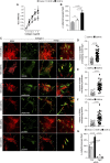
DDR1b enhances cell migration on collagen I and localizes to focal adhesions. (A) Adhesion of Vector- DDR1a-, or DDR1b-expressing HEK cells to collagen I (0–3 µg/ml). Values are the mean ± SEM of 3 independent experiments performed at least in triplicate. **p < 0.05 relative to HEK-Vector. (B) Migration of Vector- DDR1a-, or DDR1b-expressing HEK cells towards collagen I (20 μg/ml). Values are the mean ± SEM of 3 independent experiments with at least 4-6 fields/microscopic field counted. Statistical analysis in (A,B): One-way ANOVA followed by Tukey’s multiple comparison test. (C) Total internal reflection fluorescence (TIRF) microscopy of HEK-DDR1a and HEK-DDR1b cells plated on collagen I (20 µg/ml) for 1 h and then co-stained with anti-DDR1 and integrin beta 1 (12G10), FITC-phalloidin, or anti-paxillin antibodies. Overlay images reveal co-localization of DDR1b with activated integrin β1, actin or paxillin. Co-localization of DDR1a, DDR1b with 12G10 (D), actin (E), paxillin (F) at the cell membrane of cells plated on collagen I was determined using the ImageJ/JACoP analysis and the Manders’ overlap coefficient (Bolte and Cordelieres, 2006). Values are the mean ± SEM of 3-4 FAs calculated for 10-13 cells and represent the fraction of DDR1 signal overlapping with 12G10 phalloidin and paxillin. Statistical analysis: unpaired two-tailed t-test. (G) Migration of Vector- DDR1a-, and DDR1b-expressing HEK cells transfected with Cnt or Itgβ1 siRNA towards collagen I (20 μg/ml). Values are the mean ± SEM of 3 independent experiments with at least 7 fields/microscopic field counted. Statistical analysis was done with One-way ANOVA followed by Tukey’s multiple comparison test.
FIGURE 4
FIGURE 4
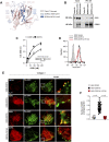
Talin binds better to DDR1b than DDR1a. (A) Model of binding between DDR1b intracellular domain (A505-Q541) and talin F3 domain. Talin F3 domain is in pink, DDR1b A505-Q541 is in light blue and the NPxY motif of DDR1b is highlighted in dark blue. Side chains of the amino acids discussed in text have been shown in sticks. (B) TurboID, DDR1a-turbo or DDR1b-turbo HEK expressing cells were plated on collagen I (30 μg/ml) for 1–3 h and then lysed and subjected to Streptavidin (SA) pull-down (PD) followed by western blotting with talin or DDR1 antibodies. (C) Immobilized talin head domain (3 µg/ml) was incubated with increasing amounts of purified cytoplasmic domains of DDR1a or DDR1b and the bound DDR1 was detected with anti-DDR1 antibody. Raw data in C were fitted with Non Linear Regression–Global Curve Fitting with one site saturation using Sigma Plot and the calculated Kd were compared using a two-tailed t-test. (D) HEK cells were stable transfected with either empty vector, DDR1b or HEK-DDR1b-Y513A cDNAs and cell populations expressing comparable levels of DDR1 were sorted by FACS. (E) Total internal reflection fluorescence (TIRF) microscopy of HEK-DDR1a, HEK-DDR1b and DDR1b-Y513A cells stable transfected with full length talin1-GFP plated on collagen I (20 µg/ml) for 1 h and then stained with anti-DDR1 or 12G10 antibody. Overlay images reveal co-localization of 12G10 and talin (positive control) and DDR1b with talin. (F) Co-localization of DDR1a, DDR1b and DDR1b-Y513A with talin1-GFP at the cell membrane of cells plated on collagen I was determined using the ImageJ/JACoP analysis and the Manders’ overlap coefficient (Bolte and Cordelieres, 2006). Values are the mean ± SEM of 3-4 FAs analyzed in more than 10 cells and represent the fraction of DDR1 signal overlapping with talin-GFP signal. Statistical analysis: one-way ANOVA followed by Tukey’s multiple comparisons test.
FIGURE 5
FIGURE 5
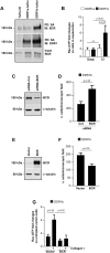
DDR1b increases cell migration by inhibiting BCR GAP activity. (A) Turbo, DDR1a-turbo or DDR1b-turbo HEK expressing cells plated on collagen I (30 μg/ml) for 1–3 h were lysed and subjected to streptavidin (SA) pulldown (PD) followed by Western blot (IB) with anti-BCR or anti-DDR1 antibodies. (B) DDR1a or DDR1b expressing cells left in suspension or plated on collagen I (30 μg/ml) for 30 min, were lysed and analyzed for the levels of active Rac. The levels of Rac-GTP were adjusted to total Rac determined by western blot analysis. Values represent fold increase relative to cells left in suspension with the lowest levels of active Rac assigned a value of 1. Values are mean ± SEM of 3 independent experiments. Statistical analysis: one-way ANOVA followed by Tukey’s multiple comparison test. (C) Western blot analysis of cells transfected with Cnt siRNA or BCR siRNA. One representative experiment of 3 independent experiments is shown. (D) Migration of DDR1b cells transfected with Cnt siRNA or BCR siRNA toward collagen I (20 μg/ml). Values are the mean ± SEM of 3 independent experiments with 7 fields/microscopic field counted. (E) Western blot analysis of cells transfected with BCR or empty vector. One representative experiment of 3 independent experiments is shown. (F) Migration of DDR1b cell transfected with BCR or empty vector towards collagen I (20 μg/ml). Values are the mean ± SEM of 3 independent experiments with 7 fields/microscopic field counted. Statistical analysis for (D,F): unpaired two-tail t-test. (G) DDR1b cells transfected with BCR or empty vector were left in suspension or plated on collagen I for 30 min, lysed and then analyzed for the levels of active Rac using a G-LISA assay as described above. Values represent fold increase relative to cells transfected with empty vector with the lowest levels of active Rac left in suspension assigned a value of 1. Values are mean ± SEM of 3 independent experiments. Statistical analysis: one-way ANOVA followed by Tukey’s multiple comparison test.
FIGURE 6
FIGURE 6
Schematic representation of collagen I-mediated DDR1a vs DDR1b function. In cells expressing DDR1a and exposed to collagen I, this receptor does not co-localize with activated integrins in focal adhesions. In contrast, in cells expressing DDR1b and exposed to collagen I, this receptor binds more efficiently to talin, co-localizes with activated integrins in focal adhesions, and enhances integrin β1-mediated cell migration. In addition, DDR1b binds the GAP BCR thus enhancing Rac1 activation and migration.

Similar articles

Cited by

References

    1. Abair T. D., Sundaramoorthy M., Chen D., Heino J., Ivaska J., Hudson B. G., et al. (2008). Cross-talk between Integrins Alpha1beta1 and Alpha2beta1 in Renal Epithelial Cells. Exp. Cel Res. 314, 3593–3604. - PMC - PubMed
    1. Alves F., Vogel W., Mossie K., Millauer B., Höfler H., Ullrich A. (1995). Distinct Structural Characteristics of Discoidin I Subfamily Receptor Tyrosine Kinases and Complementary Expression in Human Cancer. Oncogene 10, 609–618. - PubMed
    1. Ambrogio C., Gómez-López G., Falcone M., Vidal A., Nadal E., Crosetto N., et al. (2016). Combined Inhibition of DDR1 and Notch Signaling Is a Therapeutic Strategy for KRAS-Driven Lung Adenocarcinoma. Nat. Med. 22, 270–277. 10.1038/nm.4041 - DOI - PubMed
    1. An B., Abbonante V., Xu H., Gavriilidou D., Yoshizumi A., Bihan D., et al. (2016). Recombinant Collagen Engineered to Bind to Discoidin Domain Receptor Functions as a Receptor Inhibitor. J. Biol. Chem. 291, 4343–4355. 10.1074/jbc.m115.674507 - DOI - PMC - PubMed
    1. Austen K., Ringer P., Mehlich A., Chrostek-Grashoff A., Kluger C., Klingner C., et al. (2015). Extracellular Rigidity Sensing by Talin Isoform-specific Mechanical Linkages. Nat. Cel Biol. 17, 1597–1606. 10.1038/ncb3268 - DOI - PMC - PubMed