

Resveratrol prevents hypoxia-induced retinal ganglion cell death related with ErbB2
- PMID: 35310062
- PMCID: PMC8907051
- DOI: 10.18240/ijo.2022.03.04
Resveratrol prevents hypoxia-induced retinal ganglion cell death related with ErbB2
Abstract
Aim: To confirm the changes in proteins related with hypoxia-induced retinal cell death and to assess the effects of resveratrol (Res).
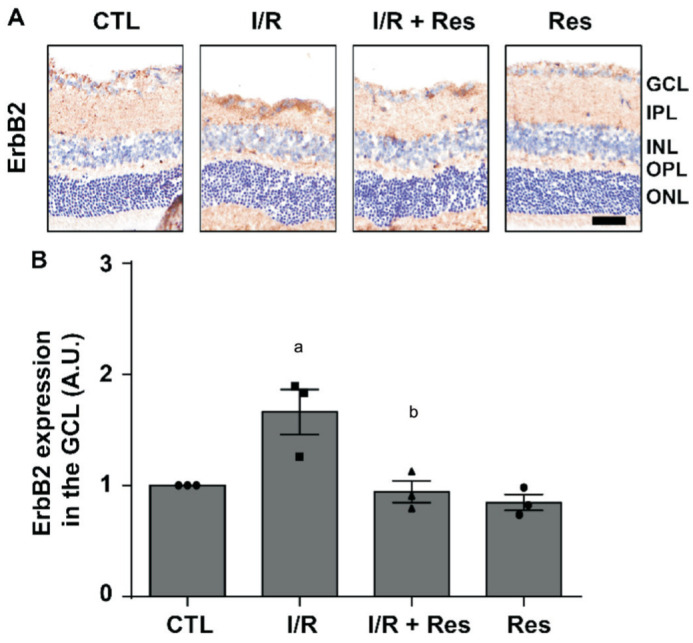
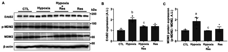
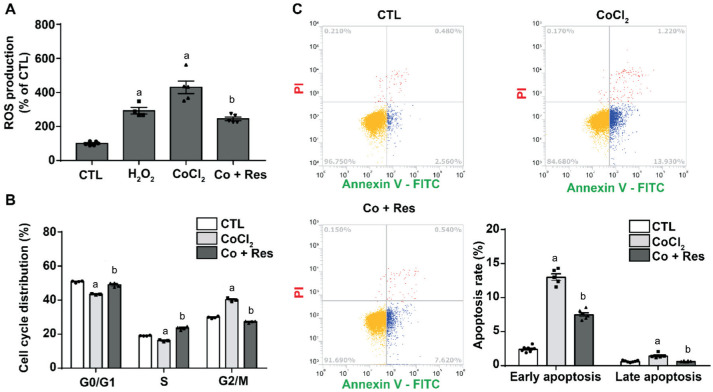
Methods: The therapeutic effect of Res was verified using an ischemic/reperfusion (I/R) model in vivo and a hypoxia modelin retinal ganglion cells (RGCs) in vitro. Death of RGCs were confirmed by TUNEL assay. Protein expression was confirmed by Western blotting and immunohistochemistry. In addition, flow cytometric analysis was used to confirm the response in the cell unit to obtain more accurate data.
Results: ErbB2 expression and apoptosis in the ganglion cell layer (GCL) increased after I/R injury. Treatment of Res rescued I/R-induced ganglion cell death, downregulated apoptosis and ErbB2 protein expression in the retina. In subsequent in vitro models, Res affects apoptosis by regulating the phosphorylation and expression of mouse double minute 2 homolog (MDM2), along with those of ErbB2. These results suggest that Res reverses GCL-specific apoptosis via downregulation of ErbB2 in ischemic injury.
Conclusion: In light of Res favorable properties, it should be evaluated in the treatment of RGC death and related retinal disease characterized by ErbB2 and MDM2 expression. Therefore, Res is appropriate therapeutic agent for treating ischemic injury-related eye diseases by targeting the expression of ErbB2 and MDM2.
Keywords: ErbB2; hypoxia; ischemia/reperfusion injury; resveratrol; retinal ganglion cell.
International Journal of Ophthalmology Press.
Figures


Similar articles
-
Resveratrol attenuates retinal ganglion cell loss in a mouse model of retinal ischemia reperfusion injury via multiple pathways.Exp Eye Res. 2021 Aug;209:108683. doi: 10.1016/j.exer.2021.108683. Epub 2021 Jun 25. Exp Eye Res. 2021. PMID: 34181937
-
Neuroprotective effects of resveratrol on retinal ganglion cells in glaucoma in rodents: A narrative review.Animal Model Exp Med. 2024 Jun;7(3):195-207. doi: 10.1002/ame2.12438. Epub 2024 May 29. Animal Model Exp Med. 2024. PMID: 38808561 Free PMC article. Review.
-
Modeling of Retina and Optic Nerve Ischemia-Reperfusion Injury through Hypoxia-Reoxygenation in Human Induced Pluripotent Stem Cell-Derived Retinal Ganglion Cells.Cells. 2024 Jan 11;13(2):130. doi: 10.3390/cells13020130. Cells. 2024. PMID: 38247823 Free PMC article.
-
Resveratrol Delays Retinal Ganglion Cell Loss and Attenuates Gliosis-Related Inflammation From Ischemia-Reperfusion Injury.Invest Ophthalmol Vis Sci. 2018 Aug 1;59(10):3879-3888. doi: 10.1167/iovs.18-23806. Invest Ophthalmol Vis Sci. 2018. PMID: 30073348
-
Resveratrol Ameliorates Retinal Ischemia/Reperfusion Injury in C57BL/6J Mice via Downregulation of Caspase-3.Curr Eye Res. 2017 Dec;42(12):1650-1658. doi: 10.1080/02713683.2017.1344713. Epub 2017 Oct 6. Curr Eye Res. 2017. PMID: 28985092
Cited by
-
Effects of Resveratrol on Vascular Function in Retinal Ischemia-Reperfusion Injury.Antioxidants (Basel). 2023 Apr 1;12(4):853. doi: 10.3390/antiox12040853. Antioxidants (Basel). 2023. PMID: 37107227 Free PMC article.
-
Etomidate protects retinal ganglion cells from hydrogen peroxide-induced injury via Nrf2/HO-1 pathway.Int J Ophthalmol. 2024 Sep 18;17(9):1606-1613. doi: 10.18240/ijo.2024.09.05. eCollection 2024. Int J Ophthalmol. 2024. PMID: 39296564 Free PMC article.
-
Glaucomatous retinal ganglion cells: death and protection.Int J Ophthalmol. 2025 Jan 18;18(1):160-167. doi: 10.18240/ijo.2025.01.20. eCollection 2025. Int J Ophthalmol. 2025. PMID: 39829615 Free PMC article. Review.
-
A comprehensive and systematic review on resveratrol supplementation as a promising candidate for the retinal disease: a focus on mechanisms of action from preclinical studies.Front Pharmacol. 2025 Jul 11;16:1615910. doi: 10.3389/fphar.2025.1615910. eCollection 2025. Front Pharmacol. 2025. PMID: 40717982 Free PMC article.
-
Resveratrol protects against diabetic retinal ganglion cell damage by activating the Nrf2 signaling pathway.Heliyon. 2024 May 6;10(9):e30786. doi: 10.1016/j.heliyon.2024.e30786. eCollection 2024 May 15. Heliyon. 2024. PMID: 38774075 Free PMC article.
References
-
- Miller JW, le Couter J, Strauss EC, Ferrara N. Vascular endothelial growth factor a in intraocular vascular disease. Ophthalmology. 2013;120(1):106–114. - PubMed
-
- Ola MS, Al-Dosari D, Alhomida AS. Role of oxidative stress in diabetic retinopathy and the beneficial effects of flavonoids. Curr Pharm Des. 2018;24(19):2180–2187. - PubMed
LinkOut - more resources
Full Text Sources
Research Materials
Miscellaneous