

Long-term outcomes of drusenoid pigment epithelium detachment in intermediate AMD treated with 577 nm subthreshold micropulse laser: a preliminary clinical study
- PMID: 35310065
- PMCID: PMC8907042
- DOI: 10.18240/ijo.2022.03.16
Long-term outcomes of drusenoid pigment epithelium detachment in intermediate AMD treated with 577 nm subthreshold micropulse laser: a preliminary clinical study
Abstract
Aim: To evaluate the long-term anatomical and visual outcomes of drusenoid pigment epithelial detachment (D-PED) in intermediate age-related macular degeneration (AMD) eyes treated with 577 nm yellow subthreshold micropulse laser (SML).
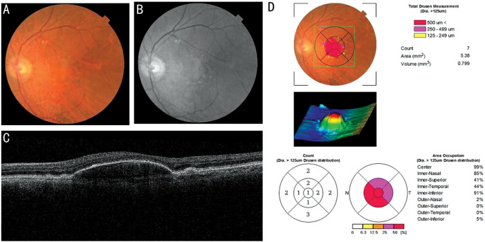
Methods: In this retrospective study, 21 eyes of 16 patients with D-PED in intermediate AMD were consecutively included and assessed. All the eyes were treated with 577 nm SML in several sessions according to D-PED growth status. The logarithm of the minimum angle of resolution (logMAR) best-corrected visual acuity (BCVA) were assessed at the initial visit and after treatment. Spectral-domain optical coherence tomography (SD-OCT) was performed to evaluate the D-PED lifecycle by volumetric calculations. Regression analysis was used to determine the breakpoint, growth, and collapse rate of the D-PED lesions. The progression to advanced AMD was also documented.
Results: All the eyes were treated with SML for 2.9±1.0 sessions. The mean follow-up period was 25.3±12.6mo. The BCVA was stable from the baseline to final visit. All the eyes were categorized into two groups according to the anatomical changes of the D-PED lesion: the collapse group (n=6, 28.6%) and non-collapse group (n=15, 71.4%). The change in logMAR BCVA did not differ significantly between the collapse group 0.00 (-0.31, 0.85) and non-collapse group 0.00 (0.00, 0.00; P=1). Regression analysis showed that the growth rate was significantly higher in the collapse group (0.090±0.095 mm3/mo) than in the non-collapse group (0.025±0.035 mm3/mo; P<0.001). One eye (4.8%) developed macular neovascularization at 11mo after SML treatment in the non-collapse group. Three eyes (14.3%) developed geographic atrophy (GA) in the collapse group.
Conclusion: Compared to the natural course of D-PED reported by previous studies, our results preliminarily show that SML can alleviate visual loss and possibility of progression to advanced AMD in eyes with D-PED in intermediate AMD. A controlled clinical trial needs to further verify the benefit of the intervention.
Keywords: age-related macular degeneration; drusenoid pigment detachment; optical coherence tomography; retinal pigment epithelium; subthreshold micropulse laser.
International Journal of Ophthalmology Press.
Figures


Similar articles
-
Long-term Choroidal Thickness Changes in Eyes With Drusenoid Pigment Epithelium Detachment.Am J Ophthalmol. 2018 Jul;191:23-33. doi: 10.1016/j.ajo.2018.03.038. Epub 2018 Apr 3. Am J Ophthalmol. 2018. PMID: 29621509
-
Associations Between Retinal Pigment Epithelium and Drusen Volume Changes During the Lifecycle of Large Drusenoid Pigment Epithelial Detachments.Invest Ophthalmol Vis Sci. 2016 Oct 1;57(13):5479-5489. doi: 10.1167/iovs.16-19816. Invest Ophthalmol Vis Sci. 2016. PMID: 27760262 Free PMC article.
-
Long-term outcomes of drusenoid retinal pigment epithelium detachment in eyes with age-related macular degeneration.Indian J Ophthalmol. 2025 Jun 1;73(6):843-846. doi: 10.4103/IJO.IJO_1966_24. Epub 2025 May 28. Indian J Ophthalmol. 2025. PMID: 40434460 Free PMC article.
-
Response of vascular pigment epithelium detachment due to age-related macular degeneration to monthly treatment with ranibizumab: the prospective, multicentre RECOVER study.Acta Ophthalmol. 2017 Nov;95(7):683-689. doi: 10.1111/aos.13359. Epub 2017 Jan 13. Acta Ophthalmol. 2017. PMID: 28084038 Clinical Trial.
-
Effect of ranibizumab on serous and vascular pigment epithelial detachments associated with exudative age-related macular degeneration.Drug Des Devel Ther. 2013 Jul 10;7:565-9. doi: 10.2147/DDDT.S46610. Print 2013. Drug Des Devel Ther. 2013. PMID: 23874084 Free PMC article.
Cited by
-
Micropulse Laser Therapy as an Integral Part of Eye Disease Management.Medicina (Kaunas). 2023 Jul 28;59(8):1388. doi: 10.3390/medicina59081388. Medicina (Kaunas). 2023. PMID: 37629677 Free PMC article. Review.
-
Subthreshold laser treatment in retinal diseases: a mini review.Graefes Arch Clin Exp Ophthalmol. 2024 Jul;262(7):2337-2344. doi: 10.1007/s00417-024-06382-4. Epub 2024 Jan 27. Graefes Arch Clin Exp Ophthalmol. 2024. PMID: 38280029 Review.
-
Yellow Subthreshold Micropulse Laser in Retinal Diseases: An In-Depth Analysis and Review of the Literature.Ophthalmol Ther. 2023 Jun;12(3):1479-1500. doi: 10.1007/s40123-023-00698-w. Epub 2023 Mar 18. Ophthalmol Ther. 2023. PMID: 36933125 Free PMC article. Review.
References
-
- Mao FF, Yang XH, Yang K, Cao XS, Cao K, Hao J, Zhang Y, Wang NL. Six-year incidence and risk factors for age-related macular degeneration in a rural Chinese population: the Handan eye study. Invest Ophthalmol Vis Sci. 2019;60(15):4966. - PubMed
-
- Mitchell P, Liew G, Gopinath B, Wong TY. Age-related macular degeneration. Lancet. 2018;392(10153):1147–1159. - PubMed
-
- Balaratnasingam C, Yannuzzi LA, Curcio CA, Morgan WH, Querques G, Capuano V, Souied E, Jung J, Freund KB. Associations between retinal pigment epithelium and drusen volume changes during the lifecycle of large drusenoid pigment epithelial detachments. Invest Ophthalmol Vis Sci. 2016;57(13):5479–5489. - PMC - PubMed
LinkOut - more resources
Full Text Sources