

Abnormal Cerebrovascular Reactivity and Functional Connectivity Caused by White Matter Hyperintensity Contribute to Cognitive Decline
- PMID: 35310084
- PMCID: PMC8930816
- DOI: 10.3389/fnins.2022.807585
Abnormal Cerebrovascular Reactivity and Functional Connectivity Caused by White Matter Hyperintensity Contribute to Cognitive Decline
Abstract
Aims: This study aimed to investigate the relationships of impaired cerebrovascular reactivity (CVR) and abnormal functional connectivity (FC) with white matter hyperintensity (WMH)-related cognitive decline.
Methods: A total of 233 WMH subjects were recruited and categorized into WMH-I (n = 106), WMH-II (n = 72), and WMH-III (n = 55) groups according to Fazekas visual rating scale. All participants underwent neuropsychological tests and multimodal MRI scans, including 3D-T1, and resting-state functional magnetic resonance imaging (rs-fMRI). The alterations of CVR maps and FC were further explored.
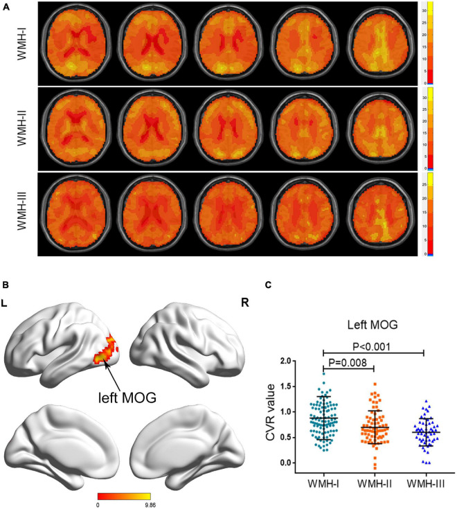
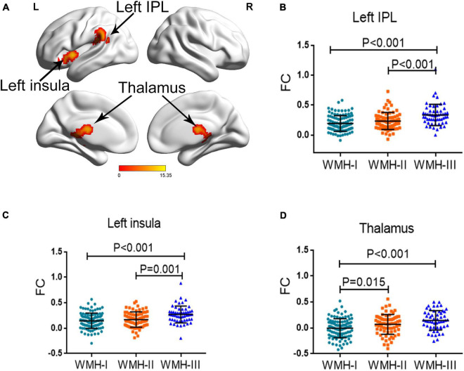
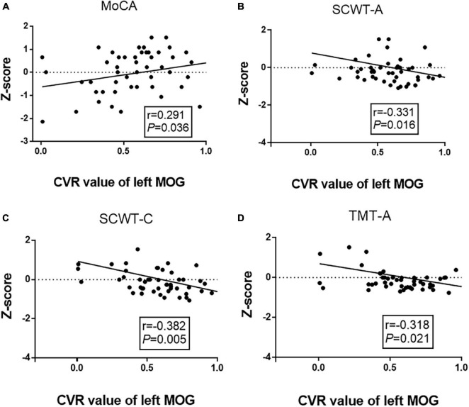
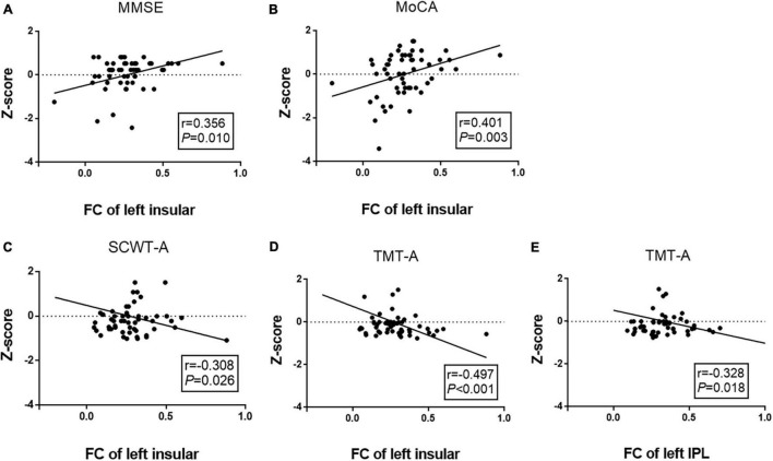
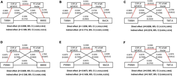
Results: Subjects with a higher WMH burden displayed a lower CVR in the left medial occipital gyrus (MOG). The FC analysis using MOG as a seed revealed that the FC of the left insula, left inferior parietal lobule, and thalamus changed abnormally as WMH aggravated. After adjusting for age, gender, and education years, the serial mediation analysis revealed that periventricular white matter hyperintensity contributes indirectly to poorer Mini-Mental State Examination (MMSE) scores (indirect effect: β = -0.1248, 95% CI: -0.4689, -0188), poorer Montreal Cognitive Assessment (MoCA) (indirect effect: β = -0.1436, 95% CI: -0.4584, -0.0292) scores, and longer trail making tests A (TMT-A) (indirect effect: β = 0.1837, 95% CI: 0.0069, 0.8273) times, specifically due to the lower CVR of the left MOG and the higher FC of the left insula-MOG.
Conclusion: The CVR decline of the left MOG and the abnormal FC of the left insula-MOG attributed to WMH progression were responsible for the poor general cognition (MMSE and MoCA) and information processing speed (TMT-A). The left MOG may act as a connection, which is involved in the processing of cognitive biases by connecting with the left insula-cortical regions in WMH individuals.
Keywords: cerebrovascular reactivity; cognitive decline; functional connectivity; rs-fMRI; white matter hyperintensity.
Copyright © 2022 Yang, Qin, Chu, Xu, Ni, Ma, Shao, Huang, Zhang, Zhang and Xu.
Conflict of interest statement
The authors declare that the research was conducted in the absence of any commercial or financial relationships that could be construed as a potential conflict of interest.
Figures
Similar articles
-
Lower Cerebrovascular Reactivity Contributed to White Matter Hyperintensity-Related Cognitive Impairment: A Resting-State Functional MRI Study.J Magn Reson Imaging. 2021 Mar;53(3):703-711. doi: 10.1002/jmri.27376. Epub 2020 Sep 29. J Magn Reson Imaging. 2021. PMID: 32996183
-
Lower cerebrovascular reactivity in prefrontal cortex and weaker negative functional connectivity between prefrontal cortex and insula contribute to white matter hyperintensity-related anxiety or depression.J Affect Disord. 2024 Jun 1;354:526-535. doi: 10.1016/j.jad.2024.03.094. Epub 2024 Mar 19. J Affect Disord. 2024. PMID: 38513774
-
Differential Effects of Confluent and Nonconfluent White Matter Hyperintensities on Functional Connectivity in Mild Cognitive Impairment.Brain Connect. 2020 Dec;10(10):547-554. doi: 10.1089/brain.2020.0784. Epub 2020 Nov 16. Brain Connect. 2020. PMID: 33050714
-
Disrupted functional and structural connectivity within default mode network contribute to WMH-related cognitive impairment.Neuroimage Clin. 2019;24:102088. doi: 10.1016/j.nicl.2019.102088. Epub 2019 Nov 12. Neuroimage Clin. 2019. PMID: 31795048 Free PMC article.
-
Tract Specific White Matter Lesion Load Affects White Matter Microstructure and Their Relationships With Functional Connectivity and Cognitive Decline.Front Aging Neurosci. 2022 Feb 2;13:760663. doi: 10.3389/fnagi.2021.760663. eCollection 2021. Front Aging Neurosci. 2022. PMID: 35185514 Free PMC article.
Cited by
-
Connectome gradient dysfunction contributes to white matter hyperintensity-related cognitive decline.CNS Neurosci Ther. 2024 Jul;30(7):e14843. doi: 10.1111/cns.14843. CNS Neurosci Ther. 2024. PMID: 38997814 Free PMC article.
-
Altered local gyrification and functional connectivity in type 2 diabetes mellitus patients with mild cognitive impairment: A pilot cross-sectional small-scale single center study.Front Aging Neurosci. 2022 Sep 20;14:934071. doi: 10.3389/fnagi.2022.934071. eCollection 2022. Front Aging Neurosci. 2022. PMID: 36204559 Free PMC article.
-
Improvement of brain perfusion in patients with chronic brain ischemia at epidural spinal cord electrical stimulation.Front Surg. 2022 Sep 23;9:1026079. doi: 10.3389/fsurg.2022.1026079. eCollection 2022. Front Surg. 2022. PMID: 36211284 Free PMC article.
-
Resting-state functional magnetic resonance imaging study on cerebrovascular reactivity changes in the precuneus of Alzheimer's disease and mild cognitive impairment patients.Sci Rep. 2025 Jan 2;15(1):363. doi: 10.1038/s41598-024-82769-x. Sci Rep. 2025. PMID: 39747269 Free PMC article.
-
Advances in differential diagnosis of cerebrovascular diseases in magnetic resonance imaging: a narrative review.Quant Imaging Med Surg. 2023 Apr 1;13(4):2712-2734. doi: 10.21037/qims-22-750. Epub 2023 Feb 22. Quant Imaging Med Surg. 2023. PMID: 37064346 Free PMC article. Review.
References
-
- Babiloni C., Del Percio C., Boccardi M., Lizio R., Lopez S., Carducci F., et al. (2015). Occipital sources of resting-state alpha rhythms are related to local gray matter density in subjects with amnesic mild cognitive impairment and Alzheimer’s disease. Neurobiol. Aging 36 556–570. 10.1016/j.neurobiolaging.2014.09.011 - DOI - PMC - PubMed
LinkOut - more resources
Full Text Sources