. 2015 Oct;116(2):377-396.
doi: 10.1111/bij.12598. Epub 2015 Sep 8.

Adaptive body patterning, three-dimensional skin morphology and camouflage measures of the slender filefish Monacanthus tuckeri on a Caribbean coral reef

Affiliations

Adaptive body patterning, three-dimensional skin morphology and camouflage measures of the slender filefish Monacanthus tuckeri on a Caribbean coral reef

Justine J Allen et al. Biol J Linn Soc Lond. 2015 Oct.

Abstract

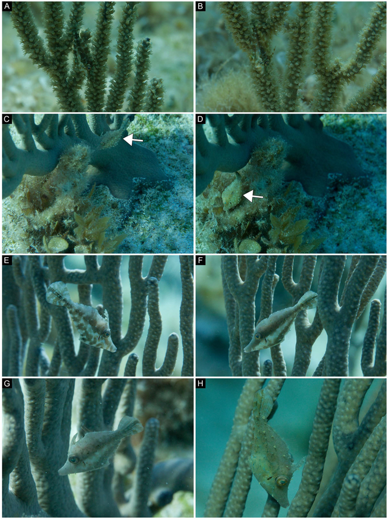
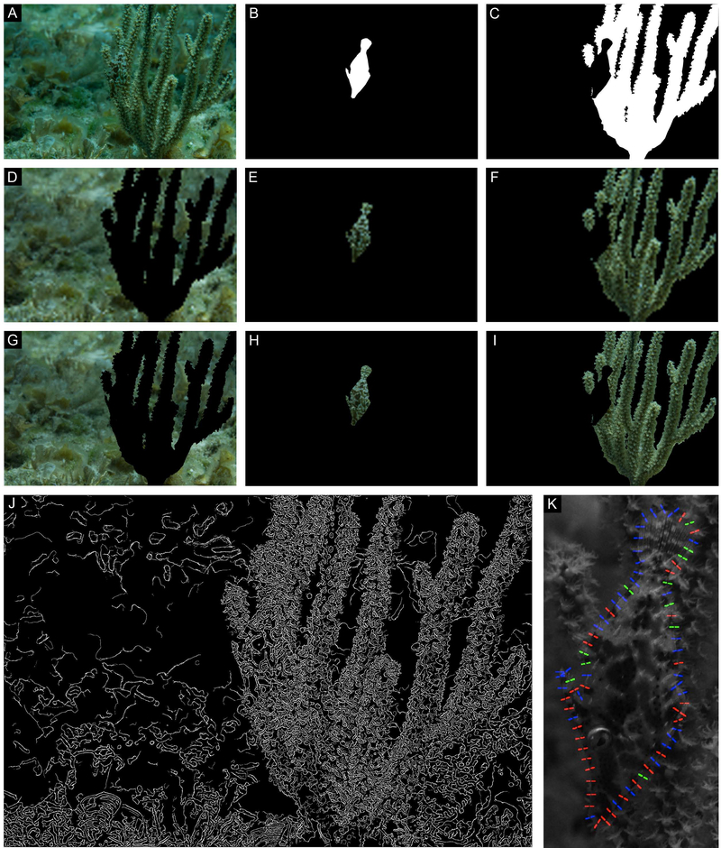
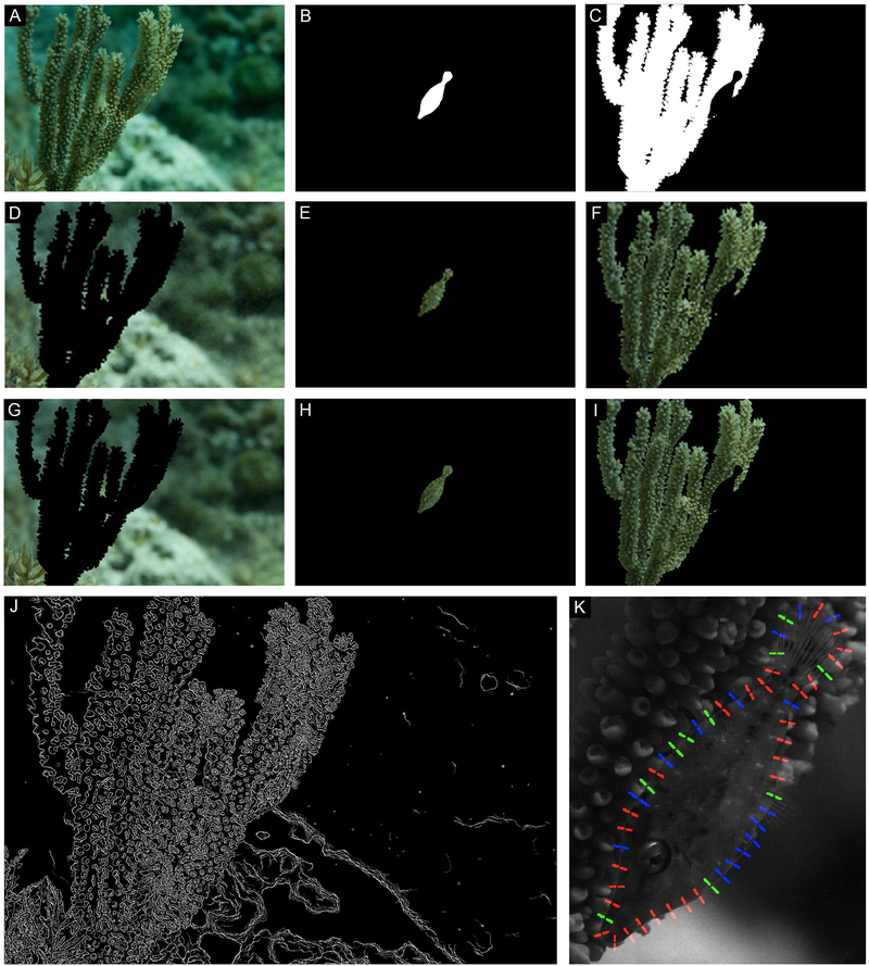
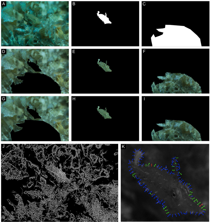
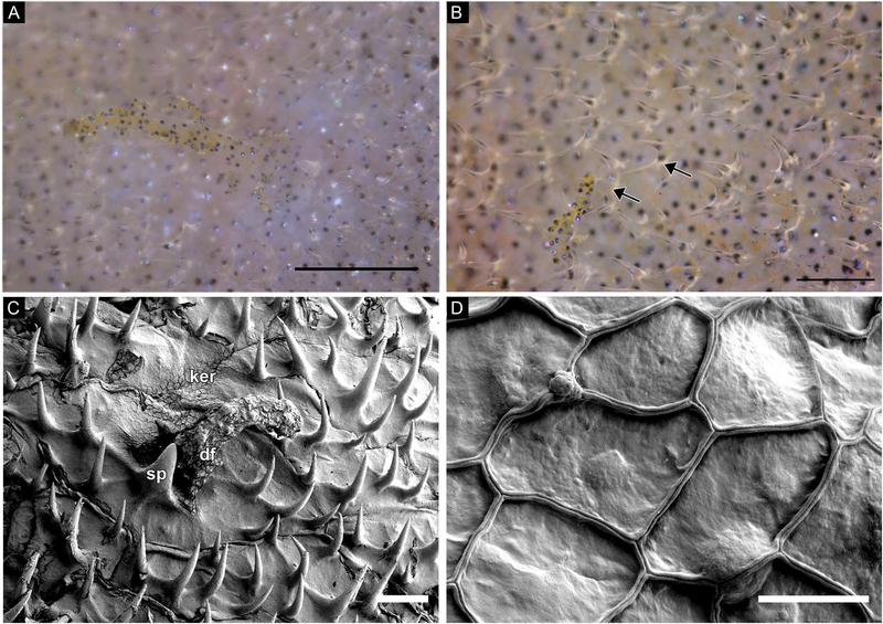
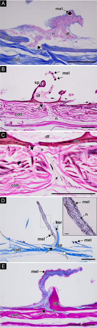
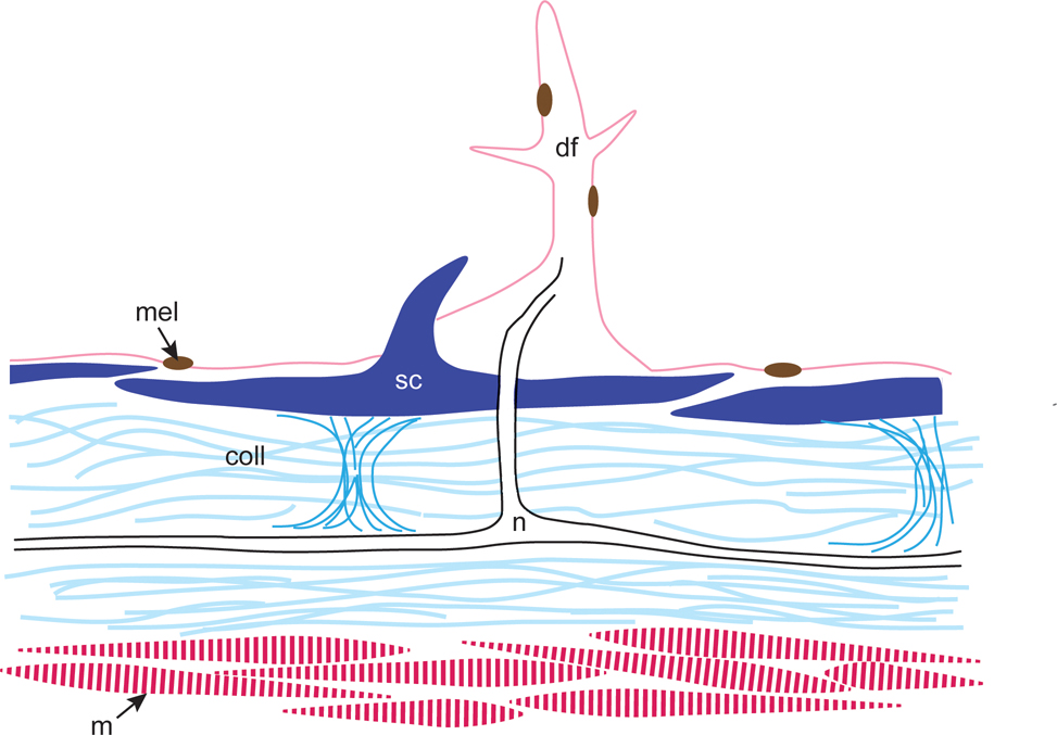
The slender filefish is a master of adaptive camouflage and can change its appearance within 1-3 seconds. Videos and photographs of this animal's cryptic body patterning and behavior were collected in situ under natural light on a Caribbean coral reef. We present an ethogram of body patterning components that includes large- and small-scale spots, stripes and bars that confer a variety of cryptic patterns amidst a range of complex backgrounds. Field images were analyzed to investigate two aspects of camouflage effectiveness: (i) the degree of color resemblance between animals and their nearby visual stimuli and (ii) the visibility of each fish's actual body outline versus its illusory outline. Most animals more closely matched the color of nearby visual stimuli than that of the surrounding background. Three-dimensional dermal flaps complement the melanophore skin patterns by enhancing the complexity of the fish's physical skin texture to disguise its actual body shape, and the morphology of these structures was studied. The results suggest that the body patterns, skin texture, postures and swimming orientations putatively hinder both the detection and recognition of the fish by potential visual predators. Overall, the rapid speed of change of multiple patterns, color blending with nearby backgrounds, and the physically complicated edge produced by dermal flaps effectively camouflage this animal among soft corals and macroalgae in the Caribbean Sea.

Keywords: cirrus; color change; coral reef ecology; cutaneous appendages; fronds; irregular marginal form; papillae; skin filaments; texture.


Figures

Figure 1:
Figure 1:
(A) Drawing illustrating gross morphology including major lateral line (dashed line), fin rays and dermal flaps. Dermal flaps exist in several sizes and are roughly drawn to scale. (B) Drawing of the 16 body pattern components identified; numbers correspond to elements identified in the text and in Table 1. These drawings illustrate the location, size, placement, and value (i.e., expressed “light” or “dark”) of all possible components for this species, identified according to our field and laboratory data, not specific patterns.
Figure 2:
Figure 2:
(A-B) Field images from an animal moving around a shelf-knob sea rod (Eunicea succinea); B was taken 11 seconds after A. (C-H) Field images from a different animal showing a variety of body patterns against different soft corals. (C-D) This filefish (white arrows) changed its body pattern when it moved from the base of a bent sea rod (Plexaura flexusa, polyps retracted) to nearby macroalgae (Sargassum sp.); D was taken 4 seconds after C. (E-G) A variety of patterns in the same animal on a single type of coral (bent sea rod, P. flexuosa); F was taken 33 seconds after E, G was taken 237 seconds after F. (H) The same animal changed its color and pattern according to a new visual background (a differently colored bent sea rod); H was taken 28 seconds after G.
Figure 3:
Figure 3:
(A-F) A single animal adjusted its body pattern according to a variety of visual backgrounds. (A) Drifting from one soft coral to the next, the animal remains cryptic against benthic white scroll algae (Padina jamaicensis) and manatee grass (Syringodium filiforme). (B) Animal camouflaging near a yellow sea whip (Pterogorgia citrina). B was taken 89 seconds after A. (C-D) Same animal showing different cryptic body patterns near different gorgonians. D was taken 150 seconds after C. (E) Same animal showing a single stripe while swimming swiftly in a horizontal body orientation over white scroll algae (P. jamaicensis). E was taken 285 seconds after D. (F) Same animal showing a striped body pattern in front of a yellow sea whip (P. citrina). Note that different body patterns can be effective against the same visual background (compare B with F). F was taken 67 seconds after E.
Figure 4:
Figure 4:
Same image as Fig. 3D. (A) Filefish camouflaged in situ. (B-C) Masks created to isolate the animal (B) from its stimulus and background (C). (D-F) Low resolution (pupil is 1 pixel) images of background (D), animal (E), and stimulus (F) analyzed for color match. (G-I) Higher resolution (pupil is 4 pixels) images of background (G), animal (H), and stimulus (I) analyzed for color match. (J) Results of the Canny edge detection analysis. (K) Results of the local contrast analysis; regions where contrast is similar (<10% different) are color coded green, regions where the animal is darker than background is red, and regions where the animal is lighter than the background are blue.
Figure 5:
Figure 5:
Same animal as Fig. S8. (A) Filefish camouflaged in situ. (B-C) Masks created to isolate the animal (B) from its stimulus and background (C). (D-F) Low resolution (pupil is 1 pixel) images of background (D), animal (E), and stimulus (F) analyzed for color match. (G-I) Higher resolution (pupil is 4 pixels) images of background (G), animal (H), and stimulus (I) analyzed for color match. (J) Results of the Canny edge detection analysis. (K) Results of the local contrast analysis; regions where contrast is similar (<10% different) are color coded green, regions where the animal is darker than background is red, and regions where the animal is lighter than the background are blue.
Figure 6:
Figure 6:
A) Filefish camouflaged in situ; animal was en route from one coral to another. (B-C) Masks created to isolate the animal (B) from its stimulus and background (C). (D-F) Low resolution (pupil is 1 pixel) images of background (D), animal (E), and stimulus (F) analyzed for color match. (G-I) Higher resolution (pupil is 4 pixels) images of background (G), animal (H), and stimulus (I) analyzed for color match. (J) Results of the Canny edge detection analysis. (K) Results of the local contrast analysis; regions where contrast is similar (<10% different) are color coded green, regions where the animal is darker than background is red, and regions where the animal is lighter than the background are blue.
Figure 7:
Figure 7:
(A-B) Freshly dead tissue; photographs of a branched dermal flaps covered with brown and yellow melanocytes and surrounded by smaller, transparent, forked spinules. (C) SEM image of a dermal flap surrounded by hook-like spinules. Squamous keratinocytes overlie the surface of the skin. Note the outlines of scales. (D) SEM image of the squamous keratinocytes. Abbreviations: dermal flap (df), keratinocytes (ker), spinule (sp). Scale bars: A-B, approx. 100μm; C, 100μm; D, 10μm.
Figure 8:
Figure 8:
Light micrographs of dermal flaps cut in cross section. (A) Mallory’s triple connective tissue stain. The dermal flap is composed of stratified cuboidal epithelial cells; note the nerve bundle passing through the scale (arrow). Epithelial cells and nerve bundle, pink; scales, blue and orange; collagen, blue; melanophores, dark brown. (B-C) Bielschowsky’s silver and Van Gieson’s stain. C is a high magnification view of the nerve bundle passing through the scale in B (arrows). Nerve bundle connects to fibers beneath the collagen layer. Epithelial cells and nerve bundle, light pink; scales, red and purple; nerves, pink and black; melanophores, dark brown/black; collagen, bright pink. (D-E) Two samples decalcified in Evans and Krajian fluid. (D) Weigert’s hematoxylin and Mallory’s 2 (orange G and aniline blue) stains. A nerve runs through the middle of the dermal flap (see inset). Nuclei of epithelial cells, dark purple; nerve bundle, light purple; scale and spinule, bright blue; collagen, bright blue. (E) Müller’s colloidal iron and Van Gieson’s stains. Epithelial cells are wrapped in mucopolysaccharide-rich connective tissue (blue); spinule, scale and collagen, bright pink. White arrow in D indicates attachment point between collagen and scale. Abbreviations: collagen (coll); dermal flap (df); keratinocyte (ker); melanophore (mel); nerve (n); scale (sc); spinule, (sp). Scale bars: 100μm; inset in D, 10μm.
Figure 9:
Figure 9:
Schematic diagram of a dermal flap in cross section showing skin elements. Scales are held in place with collagen; nerve fibers between collagen layers extend through an overlying scale to the core of the dermal flap. Light and medium blue, collagen (coll); pink, dermal flap (df), dark pink, muscle (m); brown, melanophore (mel); black, nerve tract (n); dark blue, scales (sc), center scale with spinule.

References

    1. Akkaynak D, Treibitz T, Xiao B, Gurkan UA, Allen JJ, Demirci U, Hanlon RT. 2014. Use of commercial off-the-shelf digital cameras for scientific data acquisition and scene-specific color calibration. Journal of the Optical Society of America A 31: 312–321. - PMC - PubMed
    1. Atz JW. 1951. Fishes that look like plants. Animal Kingdom 54:130–136.
    1. Bagnara JT, Matsumoto J. 2006. Comparative anatomy and physiology of pigment cells in nonmammalian tissues. In: Nordlund JJ, Boissy RE, Hearing VJ, King RA and Ortonne J-P, eds. Pigmentary System. Oxford, UK: Oxford University Press. 11–59.
    1. Bean TH. 1906. A Catalogue of the Fishes of Bermuda, With Notes on a Collection Made in 1905 for the Field Mueseum. Chicago: Field Columbian Museum.
    1. Ben-David J, Kritzer JP. 2005. Early life history and settlement of the slender filefish, Monacanthus tuckeri (Monacanthidae), at Calabash Caye, Turneffe Atoll, Belize. Environmental Biology of Fishes 73:275–282.