. 2021 Dec 2:14:15-30.
doi: 10.1016/j.bioactmat.2021.10.036. eCollection 2022 Aug.

Biodegradable magnesium fixation screw for barrier membranes used in guided bone regeneration

Affiliations

Biodegradable magnesium fixation screw for barrier membranes used in guided bone regeneration

Željka Perić Kačarević et al. Bioact Mater. .

Abstract

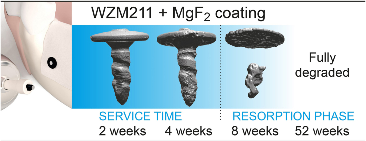
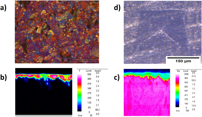
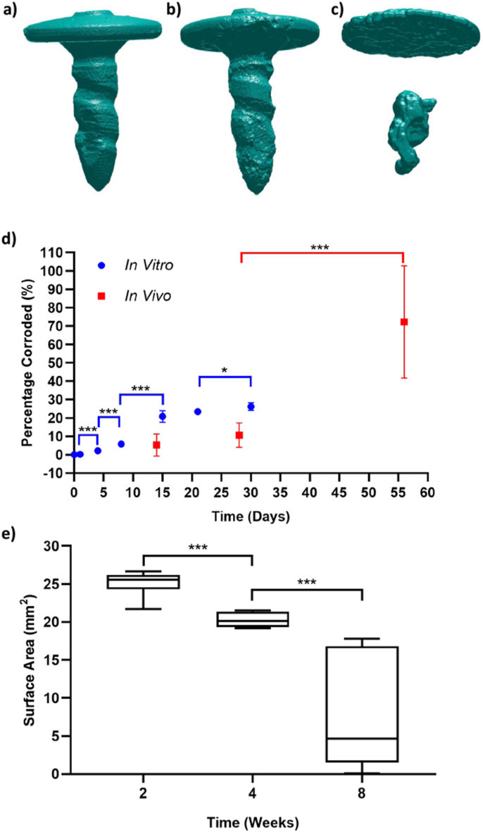
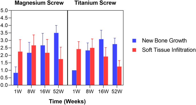
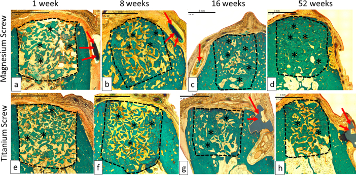
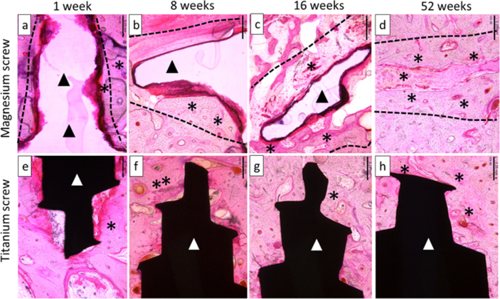
An ideal fixation system for guided bone (GBR) regeneration in oral surgery must fulfil several criteria that includes the provision of adequate mechanical fixation, complete resorption when no longer needed, complete replacement by bone, as well as be biocompatible and have a good clinical manageability. For the first time, a biodegradable magnesium fixation screw made of the magnesium alloy WZM211 with a MgF2 coating has been designed and tested to fulfill these criteria. Adequate mechanical fixation was shown for the magnesium fixation screw in several benchtop tests that directly compared the magnesium fixation screw with an equivalent polymeric resorbable device. Results demonstrated slightly superior mechanical properties of the magnesium device in comparison to the polymeric device even after 4 weeks of degradation. Biocompatibility of the magnesium fixation screw was demonstrated in several in vitro and in vivo tests. Degradation of the magnesium screw was investigated in in vitro and in vivo tests, where it was found that the screw is resorbed slowly and completely after 52 weeks, providing adequate fixation in the early critical healing phase. Overall, the magnesium fixation screw demonstrates all of the key properties required for an ideal fixation screw of membranes used in guided bone regeneration (GBR) surgeries.

Keywords: Biodegradable; Bone healing; GBR; GBR, Guided Bone Regeneration; Implant; Magnesium; Soft tissue healing.


Conflict of interest statement

The authors declare the following financial interests/personal relationships which may be considered as potential competing interests:The following authors are employees of the company biotrics bioimplants AG (Frank Witte, Marco Bartosch) and botiss biomedical AG (Zeljka Peric Kacarevic, Patrick Rider, Drazen Tadic) which companies have financed the study.A CE mark has been successfully applied for the biodegradable magnesium barrier membrane using the published data in this manuscript.

Figures

Image 1
Graphical abstract
Fig. 1
Fig. 1
a) Resorbable magnesium fixation screw (NOVAMag® fixation screw XS, botiss biomaterials GmbH) used for securing barrier membranes such as collagen, PTFE as well as pure magnesium membranes in GBR dental surgeries. b-f) Demonstrate the standard fixation protocol for the magnesium screw, in this instance for securing a collagen membrane to the mandible. b) Pilot holes with a 0.9 mm diameter are made for the fixation screw using a precision drill (NOVAMag® precision drill). c) The membrane is placed over the defect and the pilot holes located using a dental probe. d) The fixation screw is attached to a connecting device (NOVAMag® connector) and e) inserted into the pilot holes. f) Once the screw is seated, the drive of the fixation screw will shear off to leave a flat level surface to the screw head.
Fig. 2
Fig. 2
a) Schematic of the “Pull-out” test set-up according to ASTM F543. b) Schematic of the set-up used for the “shear test”. In both schematics, the grey colored pieces of equipment move, whilst the white colored pieces remain in a fixed position.
Fig. 3
Fig. 3
Approximate positions of the defect/implant sites used for the in vivo studies. a) Subperiosteally placed magnesium fixation screws in combination with a collagen membrane on the buccal side of the mandible of Yucatan minipigs. For each animal, half of the mandible was implanted with five magnesium fixation screws fixating circular sections of a collagen membrane (Bio-Gide, Geistlich). The position of each magnesium screw was marked by non-resorbable titanium screws (1.5 mm × 3 mm ProFix titanium screws, Osteogenics) The titanium screws are represented by screw heads in a diamond shape. The images b), c) and d) represent the surgical intervention performed on Beagle dogs in the in vivo performance study. b) In a preparatory surgery, four teeth between the mandibular second premolar to the first molar (PM2 to M1) on each side of the lower jaw and the corresponding teeth of the upper jaw were surgically extracted. c) After a healing period of 12 ± 2 weeks, two independent bone defects were created on each side of the lower jaw. The defects were filled with bone substitute material and covered with collagen membrane fixed with either 4 titanium or magnesium fixation screws (2 on buccal and 2 on lingual side). d) For histological evaluation, sections were taken from the central region of the defect (orange area) and from the position of the fixation screw (blue area).
Fig. 4
Fig. 4
(a) Polarized optical overview image of the Mg screw's microstructure showing. The size of the globular grains is about 6 μm with a range of 3–25 μm. (b) Fluoride mapping with WDX demonstrates that there is a continuous MgF2 coating on the screw. (c) Mg mapping with WDX confirms that Mg is mainly in the bulk material and in smaller amounts in the coating layer. (d) The Vickers hardness test indicates that there is no delamination and heavy cracks of the coating at edges of the deformation, indicating a strong adhesion of the MgF2 coating to the bulk material.
Fig. 5
Fig. 5
a-c) 3D images of the magnesium screw corroding after implantation into Yucatan minipigs after, a) 2 weeks, b) 4 weeks, and c) 8 weeks. 3D renderings were reconstructed using μCT data of explants. d) Percentage of the magnesium fixation screw corroded during immersion corrosion testing (blue circles) and after implantation in Yucatan minipigs (red squares). e) Boxplot of the mean and standard deviation for the surface area of magnesium fixation screw at the different time points of the in vivo corrosion study. In d) and e), a * indicates p < 0.05, and *** indicates p < 0.001.
Fig. 6
Fig. 6
New Bone Growth and soft issue infiltration in the central defect.
Fig. 7
Fig. 7
Representable scanned Goldner's Trichrome histology images of GBR performance study on beagles. Dotted Line = edges of the defect site; Asterisks (*) = particles of bone filler material within the defect site; Red Arrow = control and/or test articles; (a), (b), (c) and (d) are presenting a magnesium screw which is degrading over time, and by 16 weeks (c), only small residual particles of the magnesium screw are left, surrounded by new bone. The images (e), (f), (g) and (h) are presenting a control screw made of titanium, which was visible at all time points; 1 week (e), 8 weeks (f), 16 weeks (g) and 52 weeks (h). In each image, the scale bar represents 3 mm.
Fig. 8
Fig. 8
Representable scanned hemalaun eozin histology images of GBR performance study on beagles. Dotted Line = edges of the defect site where the screw was implanted; asterisks (*) = particles of new bone around screws. Black Ars = magnesium screw; (a), (b), (c) and (d) are present the magnesium screw where we can see that is degrading/reabsorbing over time. At 52 weeks (d), the magnesium screw is completely absorbed and replaced by the formation of new bone. White Ars = titanium screw; (e–h) present the titanium screw at all time points 1 week (e), 8 weeks (f), 16 weeks (g) and 52 weeks (h). In each image, the scale bar represents 0,25 mm.

References

    1. Kay S.A., Wisner-Lynch L., Marxer M., Lynch S.E. Guided bone regeneration: integration of a resorbable membrane and a bone graft material. Pract. Periodontics Aesthet. Dent. 1997;9:185—94. http://europepmc.org/abstract/MED/12698525 quiz 196. - PubMed
    1. de Rezende M.L.R., Cunha P. de O., Damante C.A., Santana A.C.P., Greghi S.L.A., Zangrando M.S.R. Cyanoacrylate adhesive as an alternative tool for membrane fixation in guided tissue regeneration. J. Contemp. Dent. Pract. 2015 doi: 10.5005/jp-journals-10024-1714. - DOI - PubMed
    1. Al-Moraissi E.A., Ellis E. Biodegradable and titanium osteosynthesis provide similar stability for orthognathic surgery. J. Oral Maxillofac. Surg. 2015;73:1795–1808. doi: 10.1016/j.joms.2015.01.035. - DOI - PubMed
    1. Cucchi A., Vignudelli E., Napolitano A., Marchetti C., Corinaldesi G. Evaluation of complication rates and vertical bone gain after guided bone regeneration with non-resorbable membranes versus titanium meshes and resorbable membranes. A randomized clinical trial. Clin. Implant Dent. Relat. Res. 2017;19:821–832. doi: 10.1111/cid.12520. - DOI - PMC - PubMed
    1. Watzinger F., Luksch J., Millesi W., Schopper C., Neugebauer J., Moser D., Ewers R. Guided bone regeneration with titanium membranes: a clinical study. Br. J. Oral Maxillofac. Surg. 2000;38:312–315. doi: 10.1054/bjom.1999.0228. - DOI - PubMed

LinkOut - more resources