Review
. 2021 Nov 30:14:234-249.
doi: 10.1016/j.bioactmat.2021.11.015. eCollection 2022 Aug.

Cells and material-based strategies for regenerative endodontics

Affiliations
Review

Cells and material-based strategies for regenerative endodontics

Zain Siddiqui et al. Bioact Mater. .

Abstract

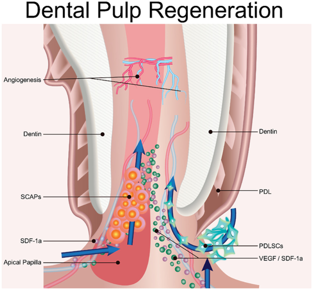
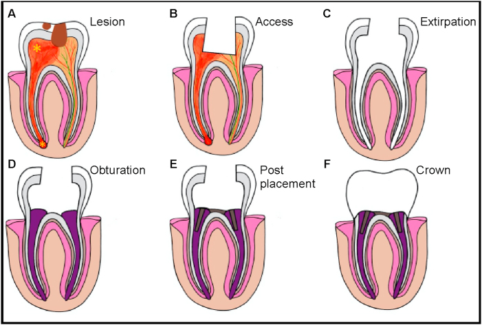
<p class = "Abstract" style = "margin: 0 cm; line-height: 32px; font-size: 12 pt; font-family: "Times New Roman", serif; color: rgb(0, 0, 0); "><span lang = "EN-US">The carious process leads to inflammation of pulp tissue. Current care options include root canal treatment or apexification. These procedures, however, result in the loss of tooth vitality, sensitivity, and healing. Pulp capping and dental pulp regeneration are continually evolving techniques to regenerate pulp tissue, avoiding necrosis and loss of vitality. Many studies have successfully employed stem/progenitor cell populations, revascularization approaches, scaffolds or material-based strategies for pulp regeneration. Here we outline advantages and disadvantages of different methods and techniques which are currently being used in the field of regenerative endodontics. We also summarize recent findings on efficacious peptide-based materials which target the dental niche.<o:p></o:p></span></p>.

Keywords: Pulp regeneration; Regenerative endodontics; Scaffolds; Stem cells; Tissue engineering.


Conflict of interest statement

V. A. K. (corresponding author) has equity interests in start-up companies attempting to translate peptides bearing angiogenic sequences. The remaining authors declare no conflicts of interest.

Figures

Image 1
Graphical abstract
Fig. 1
Fig. 1
Formation of a Tooth. Tooth development begins in utero and follows 5 stages: dental placode formation, tooth initiation, the bud stage, the cap stage and finally the bell stage. Environmental factors stimulate tooth maturation further, encasing the dental papilla beneath the enamel organ and dental follicle. Finally osteocytes foster alveolar bone formation, fibroblasts generate periodontal ligaments and cementocytes deposit cementum.
Fig. 2
Fig. 2
Endodontic Treatment of an Apical Tooth Abscess with Concurrent Caries. A) A diseased tooth with caries results in soft tissue inflammation (*) and damage to tooth enamel, exposing the pulp. Initially B) access to the pulp is obtained to C) extirpate inflamed and necrotic tissue and disinfect the tooth cavity. D) Obturation fills the emptied tooth, typically employing inert materials such as gutta-percha. Treatment is completed with E) post and core installation and finally F) placement of an artificial crown to form a protective barrier.
Fig. 3
Fig. 3
Procedure and histological analysis of pig DPSCs implanted into minipigs. A) Pig DPSCs (pDPSCs) were implanted into permanent incisors of minipigs after pulpectomy (n = 3). B) H&E staining (left) and Masson staining (right) demonstrate pulp tissue regeneration 3 months after pDPSC implantation. As a control, CH instead of pDPSCs was inserted into young permanent incisors in minipigs (n = 3). After 3 months, no pulp tissue was regenerated and only calcium hydroxide was observed. Normal pulp tissue of minipigs was stained for comparison (top). Scale bar, 50 μm. Enlarged images show odontoblasts (black arrow) and blood vessels (open arrow) in select regions of regenerated pulp tissue. Scale bar, μm. (Panel B is adapted from Xuan et al [66] (C) AAAS). Reprodcued with permission [66]. 2018, Sci. Trans. Med.
Fig. 4
Fig. 4
Schematic representation and histologic observation of a human immature permanent tooth with irreversible pulpitis after revascularization/regeneration procedure. A) Migration of cells (PDLSCs, SCAPs) and growth factors (VEGF, SDF-1a) into the tooth interior promotes angiogenesis in the tooth cavity. B) Histology of an extracted revascularized tooth, from which the MTA plug was removed prior to histological tissue processing. Connective tissue and collagen fibers fill the canal space. C) A higher magnification image of the square in B showing the apical root canal. Solid arrows indicate flattened odontoblast-like cells lining the predentin, and open arrows reveal the presence of many blood vessels filled with red blood cells. D) A higher magnification image of the rectangle in B showing the apical foramen. The presence of blood vessels is indicated by arrows. E) A higher magnification image of the square in D showing part of the root apex. Arrows indicate layers of epithelial-like HERS surrounding the root apex. Reprodcued with permission [78]. 2012, J. Endo.
Fig. 5
Fig. 5
X-ray of teeth with revascularization. A-C) Show teeth with revascularization. A) The dentinal walls (red arrows) for this patient, a 9 year old girl, are thin with a larger opening at the apex. B) after 6 months, there is calcification present at the apex. C) After 1 year, revascularization of the tooth is achieved, primarily through the bridge composed of calcium at the apical section and root lengthening. D-F) Show teeth with revascularization and PRP. D) Similar to above, the dentinal walls (red arrows) are thin with a larger opening at the apex for this patient, a 15 year old boy. This patient underwent a treatment an identical procedure however supplemented with PRP. E) At 6 months, the calcium barrier was reduced at the apex compared to the 9 year old girl and the patient reported to be symptom free. F) After 1 year, revascularization of the tooth is successful and is comparable to a normal apical tooth. Reprodcued with permission [44]. 2021, J. Endo.

Similar articles

Cited by

References

    1. Hand R.A., Frank M.E. Fundamentals of Oral Histology and Physiology. first ed. ed. John Wiley & Sons, Inc; 2015.
    1. Shchetinin E.V. Медицинский вестник Северного Кавказа; 2015. Pathogenetic Aspects of Dental Pulp Pathology.
    1. Schuurs A. John Eiley & Sons, Inc; 2012. Pathology of the Hard Dental Tissues.
    1. Bjorndal L., Mjor I.A. Pulp-dentin biology in restorative dentistry. Part 4: dental caries--characteristics of lesions and pulpal reactions. Quintessence Int. 2001;32(9):717–736. - PubMed
    1. Zaleckiene V., Peciuliene V., Brukiene V., Drukteinis S. Traumatic dental injuries: etiology, prevalence and possible outcomes. Stomatol. 2014;16(1):7–14. - PubMed