

Root and Canal Morphology of Mandibular Premolars in a Saudi Subpopulation: A Cone-Beam Computed Tomography Study
- PMID: 35310460
- PMCID: PMC8924601
- DOI: 10.1155/2022/4038909
Root and Canal Morphology of Mandibular Premolars in a Saudi Subpopulation: A Cone-Beam Computed Tomography Study
Abstract
Objectives: The efficacy of root canal therapy is dependent on a thorough understanding of both normal and aberrant root canal morphology. As a result, the purpose of this study was to use CBCT to characterize the exact root and canal morphology of mandibular premolars in a Saudi subpopulation.
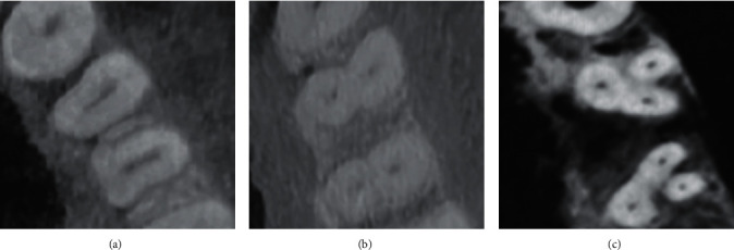
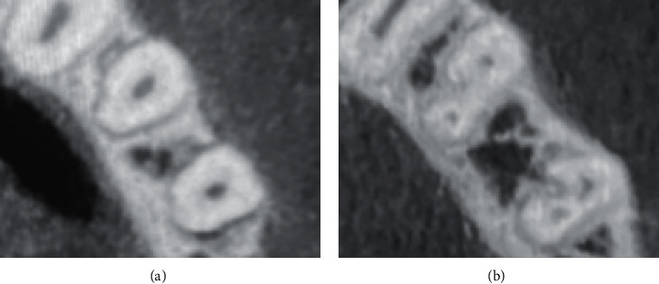
Methods: The current study included 1000 mandibular premolars (507 first premolars and 493 second premolars) with completely developed roots. CBCT was performed to assess the shape of the roots and to classify the canal anatomy according to Vertucci's classification. The incidence and similarity of the left and right sides, as well as men and women, were investigated. The data were examined using the chi-square test.
Results: Of the 507 mandibular first premolars analyzed, 484 (95.5%) had one root, whereas 23 (4.5%) had two roots. Of the 493 mandibular second premolars analyzed, 489 (99.2%) had one root, whereas four teeth had two roots (0.8%). There were no statistically significant variations in the number of roots identified across groups (p > 0.05). The most prevalent in mandibular first premolars was type I, accounting for 70.0% (n = 355) of the studied sample, followed by type II (14.2%, n = 72) and type IV (10.1%, n = 51). For mandibular second premolar, type I had the highest incidence (449 (91.1%)), followed by type II (5.7%, n = 28).
Conclusion: In a Saudi subpopulation, the majority of mandibular first and second premolar teeth had a single root with a type I canal system. On the other hand, numerous roots with various canal classifications were found.
Copyright © 2022 Saad M. Al-Zubaidi et al.
Conflict of interest statement
The authors state that they have no conflicts of interest.
Figures
Similar articles
-
Evaluation of root and canal morphology of mandibular premolar amongst Saudi subpopulation using the new system of classification: a CBCT study.BMC Oral Health. 2023 May 15;23(1):291. doi: 10.1186/s12903-023-03002-1. BMC Oral Health. 2023. PMID: 37189077 Free PMC article.
-
Assessment of root morphology and canal configuration of maxillary premolars in a Saudi subpopulation: a cone-beam computed tomographic study.BMC Oral Health. 2021 Aug 13;21(1):397. doi: 10.1186/s12903-021-01739-1. BMC Oral Health. 2021. PMID: 34389013 Free PMC article.
-
Root and Canal Morphology of Mandibular Premolar Teeth in a Kuwaiti Subpopulation: A CBCT Clinical Study.Eur Endod J. 2020 Dec;5(3):248-256. doi: 10.14744/eej.2020.40085. Eur Endod J. 2020. PMID: 33353914 Free PMC article.
-
Root Morphology and Canal Configuration of Permanent Canines Among Saudi Population: Systematic Review and Comparison with Worldwide Studies.Int J Gen Med. 2022 Aug 29;15:6849-6860. doi: 10.2147/IJGM.S380084. eCollection 2022. Int J Gen Med. 2022. PMID: 36061964 Free PMC article. Review.
-
Evidence of Second Canal between Permanent Mandibular Central and Lateral Incisors in China; a Systematic Review on CBCT Studies.Int J Dent. 2020 Dec 3;2020:8849609. doi: 10.1155/2020/8849609. eCollection 2020. Int J Dent. 2020. PMID: 33343667 Free PMC article. Review.
Cited by
-
Evaluation of root canal morphology of mandibular premolars in Pakistani population using the new classification: a CBCT study.BMC Oral Health. 2024 Nov 20;24(1):1414. doi: 10.1186/s12903-024-05149-x. BMC Oral Health. 2024. PMID: 39568006 Free PMC article.
-
Evaluation of root and canal morphology of mandibular premolar amongst Saudi subpopulation using the new system of classification: a CBCT study.BMC Oral Health. 2023 May 15;23(1):291. doi: 10.1186/s12903-023-03002-1. BMC Oral Health. 2023. PMID: 37189077 Free PMC article.
-
The importance of cone-beam computed tomography in endodontic therapy: A review.Saudi Dent J. 2023 Nov;35(7):780-784. doi: 10.1016/j.sdentj.2023.07.005. Epub 2023 Jul 7. Saudi Dent J. 2023. PMID: 38025595 Free PMC article. Review.
-
Evaluation of Root Canal Morphology of Mandibular Premolars Using Cone-beam Computed Tomography in Golestan Province, North of Iran.Iran Endod J. 2024;19(3):183-188. doi: 10.22037/iej.v19i3.40052. Iran Endod J. 2024. PMID: 39086715 Free PMC article.
-
Prevalence of Second Root and Root Canal in Mandibular and Maxillary Premolars Based on Two Classification Systems in Sub-Population of Northern Region (Saudi Arabia) Assessed Using Cone Beam Computed Tomography (CBCT): A Retrospective Study.Diagnostics (Basel). 2023 Jan 29;13(3):498. doi: 10.3390/diagnostics13030498. Diagnostics (Basel). 2023. PMID: 36766603 Free PMC article.
References
-
- Filpo–Perez C., Bramante C. M., Villas-Boas M. H., Duarte M. A. H., Versiani M. A., Ordinola-Zapata R. Micro–computed tomographic analysis of the root canal morphology of the distal root of mandibular first molar. Journal of Endodontics . 2015;41(2):231–236. doi: 10.1016/j.joen.2014.09.024. - DOI - PubMed
-
- Tahmasbi M., Jalali P., Nair M. K., Barghan S., Nair U. P., Nair U. P. Prevalence of middle mesial canals and isthmi in the mesial root of mandibular molars: an in vivo cone-beam computed tomographic study. Journal of Endodontics . 2017;43(7):1080–1083. doi: 10.1016/j.joen.2017.02.008. - DOI - PubMed
LinkOut - more resources
Full Text Sources
Research Materials
