Can super-resolution microscopy become a standard characterization technique for materials chemistry?
- PMID: 35310478
- PMCID: PMC8864713
- DOI: 10.1039/d1sc05506b
Can super-resolution microscopy become a standard characterization technique for materials chemistry?
Abstract
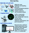
The characterization of newly synthesized materials is a cornerstone of all chemistry and nanotechnology laboratories. For this purpose, a wide array of analytical techniques have been standardized and are used routinely by laboratories across the globe. With these methods we can understand the structure, dynamics and function of novel molecular architectures and their relations with the desired performance, guiding the development of the next generation of materials. Moreover, one of the challenges in materials chemistry is the lack of reproducibility due to improper publishing of the sample preparation protocol. In this context, the recent adoption of the reporting standard MIRIBEL (Minimum Information Reporting in Bio-Nano Experimental Literature) for material characterization and details of experimental protocols aims to provide complete, reproducible and reliable sample preparation for the scientific community. Thus, MIRIBEL should be immediately adopted in publications by scientific journals to overcome this challenge. Besides current standard spectroscopy and microscopy techniques, there is a constant development of novel technologies that aim to help chemists unveil the structure of complex materials. Among them super-resolution microscopy (SRM), an optical technique that bypasses the diffraction limit of light, has facilitated the study of synthetic materials with multicolor ability and minimal invasiveness at nanometric resolution. Although still in its infancy, the potential of SRM to unveil the structure, dynamics and function of complex synthetic architectures has been highlighted in pioneering reports during the last few years. Currently, SRM is a sophisticated technique with many challenges in sample preparation, data analysis, environmental control and automation, and moreover the instrumentation is still expensive. Therefore, SRM is currently limited to expert users and is not implemented in characterization routines. This perspective discusses the potential of SRM to transition from a niche technique to a standard routine method for material characterization. We propose a roadmap for the necessary developments required for this purpose based on a collaborative effort from scientists and engineers across disciplines.
This journal is © The Royal Society of Chemistry.
Conflict of interest statement
There are no conflicts to declare.
Figures


Similar articles
-
The power of super-resolution microscopy in modern biomedical science.Adv Colloid Interface Sci. 2023 Apr;314:102880. doi: 10.1016/j.cis.2023.102880. Epub 2023 Mar 13. Adv Colloid Interface Sci. 2023. PMID: 36965225 Review.
-
The future of Cochrane Neonatal.Early Hum Dev. 2020 Nov;150:105191. doi: 10.1016/j.earlhumdev.2020.105191. Epub 2020 Sep 12. Early Hum Dev. 2020. PMID: 33036834
-
Proceedings of the Second Workshop on Theory meets Industry (Erwin-Schrödinger-Institute (ESI), Vienna, Austria, 12-14 June 2007).J Phys Condens Matter. 2008 Feb 13;20(6):060301. doi: 10.1088/0953-8984/20/06/060301. Epub 2008 Jan 24. J Phys Condens Matter. 2008. PMID: 21693862
-
Super-Resolution Microscopy as a Versatile Tool in Probing Molecular Assembly.Int J Mol Sci. 2024 Oct 26;25(21):11497. doi: 10.3390/ijms252111497. Int J Mol Sci. 2024. PMID: 39519049 Free PMC article. Review.
-
Spectrally Resolved and Functional Super-resolution Microscopy via Ultrahigh-Throughput Single-Molecule Spectroscopy.Acc Chem Res. 2018 Mar 20;51(3):697-705. doi: 10.1021/acs.accounts.7b00545. Epub 2018 Feb 14. Acc Chem Res. 2018. PMID: 29443498
Cited by
-
Unveiling nanoparticle-immune interactions: how super-resolution imaging illuminates the invisible.Nanoscale. 2025 Jan 16;17(3):1213-1224. doi: 10.1039/d4nr03838j. Nanoscale. 2025. PMID: 39618290 Review.
-
Visualizing the Heterogeneity in Homogeneous Supramolecular Polymers.J Am Chem Soc. 2024 Jul 24;146(29):19974-19985. doi: 10.1021/jacs.4c03562. Epub 2024 Jul 10. J Am Chem Soc. 2024. PMID: 38986035 Free PMC article.
-
Beyond DNA: new probes for PAINT super-resolution microscopy.Chem Commun (Camb). 2023 Jul 4;59(54):8332-8342. doi: 10.1039/d3cc00757j. Chem Commun (Camb). 2023. PMID: 37306078 Free PMC article. Review.
-
Probing Peptide Assembly and Interaction via High-Resolution Imaging Techniques: A Mini Review.Int J Mol Sci. 2025 Apr 23;26(9):3998. doi: 10.3390/ijms26093998. Int J Mol Sci. 2025. PMID: 40362238 Free PMC article. Review.
-
Unsupervised Data-Driven Reconstruction of Molecular Motifs in Simple to Complex Dynamic Micelles.J Phys Chem B. 2023 Mar 23;127(11):2595-2608. doi: 10.1021/acs.jpcb.2c08726. Epub 2023 Mar 8. J Phys Chem B. 2023. PMID: 36891625 Free PMC article.
References
-
- Pujals S. Feiner-Gracia N. Delcanale P. Voets I. Albertazzi L. Nat. Rev. Chem. 2019;3:68–84. doi: 10.1038/s41570-018-0070-2. - DOI
Publication types
LinkOut - more resources
Full Text Sources

