Case Reports
. 2021 Jul 5;2(1):e13.
doi: 10.1002/deo2.13. eCollection 2022 Apr.

Serrated polyposis syndrome with multiple inverted lesions in the colon: Case report and elucidation of morphogenetic mechanism

Affiliations
Case Reports

Serrated polyposis syndrome with multiple inverted lesions in the colon: Case report and elucidation of morphogenetic mechanism

Chihiro Yoshikawa et al. DEN Open. .

Abstract

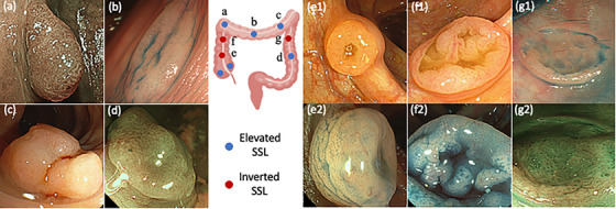
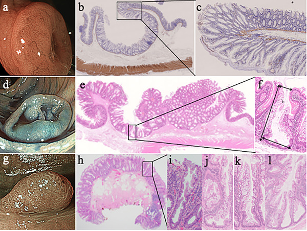
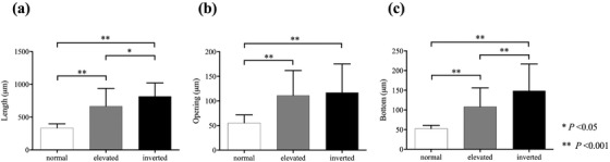
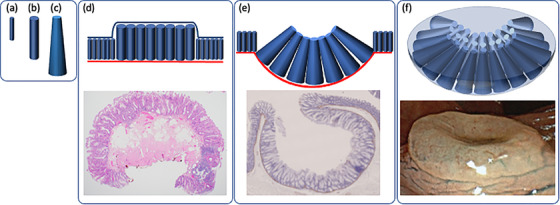
A 70-year-old man underwent surveillance colonoscopy following surgery for occlusive sigmoid colon cancer. The procedure revealed nine sessile serrated lesions (SSLs), including three inverted lesions. Endoscopic and surgical resections were performed. All nine lesions were confirmed pathologically as SSL, and the patient was diagnosed with serrated polyposis syndrome (SPS). Three inverted SSLs (iSSLs) showed endophytic growth without epithelial misplacement. Crypt analysis revealed that iSSL crypts were wider at the bottom than the opening, roughly resembling a frustoconical shape. Our results suggest that a horizontal arrangement of frustoconical crypts leads to hemispherical deformation of the muscularis mucosa, forming an inverted shape. This is the first report to reveal the morphogenesis of iSSLs from the shape of the crypt.

Keywords: depressed surface; endophytic growth; frustoconical crypt; inverted sessile serrated lesions; serrated polyposis syndrome.


Conflict of interest statement

The authors declare no conflicts of interest.

Figures

FIGURE 1
FIGURE 1
Distribution of nine SSLs in the colon and their colonoscopic appearances. Colonoscopic images are shown for all SSLs except two eSSLs in the ascending colon, which were not detected preoperatively and therefore no colonoscopic images are available. Endoscopic images of eSSLs (a–d) and iSSLs (e–g) are shown. White light appearance (e1) and indigo‐carmine dye splaying image (e2) of an iSSL 10 mm in diameter in the ascending. White light appearance (f1) and indigo‐carmine dye splaying image (f2) of an iSSL 18 mm in diameter in the ascending colon. Indigo carmine dye splaying image (g1) and blue laser imaging (g2) of iSSL in the descending colon
FIGURE 2
FIGURE 2
Histological images of iSSLs and eSSLs. (a) Blue laser imaging of an iSSL 10 mm in diameter in the ascending colon. (b) Histopathologic image of this iSSL. The proper muscular layer is preserved. The depressed portions comprise serrated crypts with inverted growth, and the marginal elevations comprise normal crypts (Desmin stain, 2 ×). (c) High‐magnification image of a bend section. The muscularis mucosa is bent to form circumferential ridges at the boundary between the serrated crypts and the surrounding normal crypts (Desmin stain, 2 ×). (d) Indigo‐carmine dye splaying image of an iSSL 18 mm in diameter, with minute elevations seen inside the central wide depression in the ascending colon. (e) The lesions have depressed areas with endophytic growth. Crypt misplacement is not observed (H&E stain, 2 ×). (f) Enlarged image of a crypt from a depressed part of the iSSL. The bottom of the crypt was wider than the opening, and crypt length was greater than that of normal crypts. We measured arrows as crypt length and the widths of crypt openings and bottoms. (g) Colonoscopic image of an eSSL. (h) Histopathologic image of eSSL (H&E stain, 2 ×). (i) Enlarged image of a crypt composed of eSSLs. The widths of the bottom and the opening are the same. (j) Histopathologic image of L‐shaped crypt in the 18‐mm diameter iSSL. (k) Histopathologic image of inverted T‐shaped crypt in the 18‐mm diameter iSSL. (l) Histopathologic image of irregularly branching crypts in the 18‐mm diameter iSSL.
FIGURE 3
FIGURE 3
The results of crypt analysis of normal mucosa, eSSLs, and iSSLs. Statistical differences in crypt length (a), opening (b), and bottom (c) between normal mucosa, eSSLs, and iSSLs were analyzed with one‐way ANOVA and Tukey's HSD post hoc test. (a) Mean crypt length in normal mucosa, eSSLs, and iSSLs was 335.7 ± 62.0, 668.7 ± 269.4, and 817.7 ± 204.6, respectively. (b) Mean width of the crypt opening in normal mucosa, eSSLs, and iSSLs was 55.3 ± 16.5, 111.3 ± 50.5, and 117.0 ± 58.4, respectively. (c) Mean width of the crypt bottom in normal mucosa, eSSLs, and iSSLs was 52.6 ± 8.2, 108.3 ± 47.7, and 148.6 ± 68.2, respectively.
FIGURE 4
FIGURE 4
Graphic representation of crypt types and the morphogenetic mechanism. (a) Normal, (b) eSSL, and (c) iSSL crypts. (d) Image showing crypt arrangement in an eSSL. The elevated portions consist of large columnar crypts. (e) Image showing crypt arrangement in an iSSL. The depressed portions consist of frustoconical crypts, and the marginal elevations contain columnar shaped crypts. The red line indicates the muscularis mucosa, which bends to form a circumferential ridge at the boundary between the columnar crypts and surrounding normal crypts. (f) 3D image of an iSSL. The frustoconical crypts grow in a circular arrangement, compressing the muscularis mucosa convexly toward the submucosal layer, to form a hemispherical surface.

Similar articles

Cited by

References

    1. Dekker E, Bleijenberg A, Balaguer F. Update on the world Health organization criteria for diagnosis of serrated polyposis syndrome. Gastroenterology 2020; 158: 1520–3. - PubMed
    1. Uraoka T, Saito Y, Ikematsu H, Yamamoto K, Sano Y. Sano's capillary pattern classification for narrow‐band imaging of early colorectal lesions. Dig Endosc 2011; 23 (Suppl 1): 112–5. - PubMed
    1. Kimura T, Yamamoto E, Yamano HO, et al. A novel pit pattern identifies the precursor of colorectal cancer derived from sessile serrated adenoma. Am J Gastroenterol 2012; 107: 460–9. - PubMed
    1. Suzuki D, Matsumoto S, Mashima H. A case with serrated polyposis syndrome controlled by multiple applications of endoscopic mucosal resection and endoscopic submucosal dissection. Am J Case Rep 2017; 18: 304–7. - PMC - PubMed
    1. Stepherd NA. Inverted hyperplastic polyposis of the colon. J Clin Pathol 1993; 46: 56–60. - PMC - PubMed

Publication types