

Visibility of early gastric cancer in texture and color enhancement imaging
- PMID: 35310718
- PMCID: PMC8828244
- DOI: 10.1002/deo2.46
Visibility of early gastric cancer in texture and color enhancement imaging
Abstract
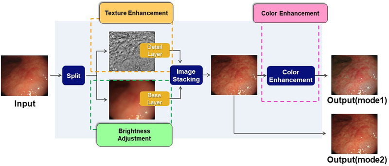
Objective: There are little data regarding the efficacy of texture and color enhancement imaging (TXI) for early gastric cancer (EGC) diagnosis. This study aimed to compare the color difference and visibility of EGC between white light imaging (WLI) and TXI.
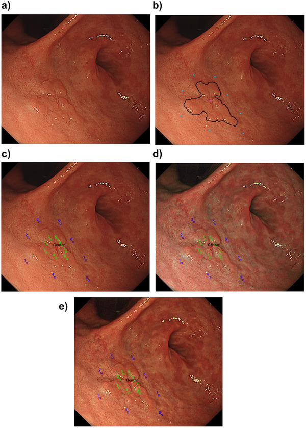
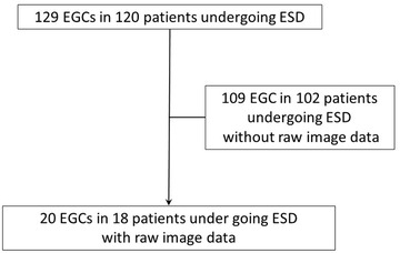
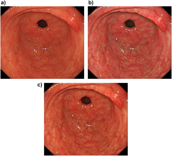
Methods: This study included 20 EGCs of 18 patients undergoing endoscopic submucosal dissection. Still images of EGC in WLI, TXI mode 1 (with color enhancement), and TXI mode 2 (without color enhancement), which were consistent in distance, angle, and air insufflation, were constructed by computer simulation. The center of the lesion, eight equal peripheral points 5 mm outside the lesion, and eight inner points two-thirds of the distance from peripheral points to the EGC lesion center were annotated. Mean color differences (ΔE) of the area between peripheral and inner points per lesion in WLI, TXI mode 1, and TXI mode 2 were analyzed. In addition, four endoscopists independently scored the visibility of EGC images of TXI mode 1 and 2 compared with WLI.
Results: Clinicopathological characteristics were as follows: 0-IIa/0-IIb/0-IIc/0-IIa+IIc = 6/1/11/2, reddish/pale = 10/10, differentiated/undifferentiated = 18/2, median tumor size = 13.5 mm. Mean ΔE ± SD = WLI/TXI mode1/TXI mode2 = 10.3 ± 4.7, 15.5 ± 7.8, and 12.7 ± 6.1, respectively. Mean ΔE was significantly higher in TXI mode 1 than in WLI. Visibility (improved/no change/decreased) was 7/13/0 and 4/16/0 in TXI mode 1 and 2, respectively. The visibility was significantly more commonly improved in the macroscopic type 0-IIc or 0-IIb than in 0-IIa or IIa+IIc in TXI mode 1.
Conclusions: TXI could improve the visibility of EGC compared with WLI.
Keywords: early gastric cancer; image enhanced endoscopy; texture and color enhancement imaging; visibility.
© 2021 The Authors. DEN Open published by John Wiley & Sons Australia, Ltd on behalf of Japan Gastroenterological Endoscopy Society.
Conflict of interest statement
The author Seiichiro Abe was supported by a research grant from Olympus Medical Systems (C2020‐036). The authors Seiichiro Abe and Yutaka Saito received honoraria for lectures from Olympus Medical Systems. The author Seiichiro Abe is an associate editor of DEN Open.
Figures
Similar articles
-
Visibility evaluation of colorectal lesion using texture and color enhancement imaging with video.DEN Open. 2022 Jan 11;2(1):e90. doi: 10.1002/deo2.90. eCollection 2022 Apr. DEN Open. 2022. PMID: 35310754 Free PMC article.
-
Using texture and colour enhancement imaging to evaluate gastrointestinal diseases in clinical practice: a review.Ann Med. 2022 Dec;54(1):3315-3332. doi: 10.1080/07853890.2022.2147992. Ann Med. 2022. PMID: 36420822 Free PMC article. Review.
-
Texture and Color Enhancement Imaging Increases Color Changes and Improves Visibility for Squamous Cell Carcinoma Suspicious Lesions in the Pharynx and Esophagus.Diagnostics (Basel). 2021 Oct 23;11(11):1971. doi: 10.3390/diagnostics11111971. Diagnostics (Basel). 2021. PMID: 34829318 Free PMC article.
-
Visibility of early gastric cancers by texture and color enhancement imaging using a high-definition ultrathin transnasal endoscope.Sci Rep. 2023 Feb 3;13(1):1994. doi: 10.1038/s41598-023-29284-7. Sci Rep. 2023. PMID: 36737509 Free PMC article.
-
Analysis of Texture and Color Enhancement Imaging for Improving the Visibility of Non-polypoid Colorectal Lesions.Dig Dis Sci. 2022 Dec;67(12):5657-5665. doi: 10.1007/s10620-022-07460-5. Epub 2022 Mar 22. Dig Dis Sci. 2022. PMID: 35318554 Review.
Cited by
-
Efficacy of texture and color enhancement imaging for the visibility and diagnostic accuracy of non-polypoid colorectal lesions.DEN Open. 2024 May 29;5(1):e380. doi: 10.1002/deo2.380. eCollection 2025 Apr. DEN Open. 2024. PMID: 38817687 Free PMC article.
-
Visibility evaluation of colorectal lesion using texture and color enhancement imaging with video.DEN Open. 2022 Jan 11;2(1):e90. doi: 10.1002/deo2.90. eCollection 2022 Apr. DEN Open. 2022. PMID: 35310754 Free PMC article.
-
Third-Generation High-Vision Ultrathin Endoscopy Using Texture and Color Enhancement Imaging and Narrow-Band Imaging to Evaluate Barrett's Esophagus.Diagnostics (Basel). 2022 Dec 13;12(12):3149. doi: 10.3390/diagnostics12123149. Diagnostics (Basel). 2022. PMID: 36553156 Free PMC article.
-
Image-enhanced endoscopy in upper gastrointestinal disease: focusing on texture and color enhancement imaging and red dichromatic imaging.Clin Endosc. 2025 Mar;58(2):163-180. doi: 10.5946/ce.2024.159. Epub 2024 Nov 6. Clin Endosc. 2025. PMID: 39722144 Free PMC article. Review.
-
Using texture and colour enhancement imaging to evaluate gastrointestinal diseases in clinical practice: a review.Ann Med. 2022 Dec;54(1):3315-3332. doi: 10.1080/07853890.2022.2147992. Ann Med. 2022. PMID: 36420822 Free PMC article. Review.
References
-
- Ferlay J, Colombet M, Soerjomataram I, et al. Estimating the global cancer incidence and mortality in 2018: GLOBOCAN sources and methods. Int J Cancer 2018;. 144: 1941–53. - PubMed
-
- Katai H, Ishikawa T, Akazawa K, et al. Five‐year survival analysis of surgically resected gastric cancer cases in Japan: A retrospective analysis of more than 100,000 patients from the nationwide registry of the Japanese Gastric Cancer Association (2001‐2007). Gastric Cancer 2018; 21: 144–54. - PubMed
-
- Hamashima C. Update version of the Japanese guidelines for gastric cancer screening. Jpn J Clin Oncol 2018; 48: 673–83. - PubMed
-
- Jun JK, Choi KS, Lee HY, et al. Effectiveness of the Korean national cancer screening program in reducing gastric cancer mortality. Gastroenterology 2017; 152: 1319–28. - PubMed
-
- Amin A, Gilmour H, Graham L, Paterson‐Brown S, Terrace J, Crofts TJ. Gastric adenocarcinoma missed at endoscopy. J R Coll Surg Edinb 2002; 47: 681–4. - PubMed
LinkOut - more resources
Full Text Sources