

The Potential of Photoacoustic Imaging in Radiation Oncology
- PMID: 35311156
- PMCID: PMC8928467
- DOI: 10.3389/fonc.2022.803777
The Potential of Photoacoustic Imaging in Radiation Oncology
Abstract
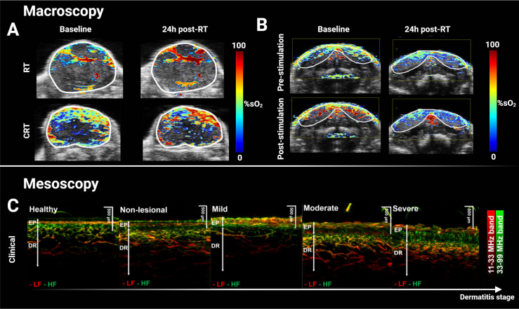
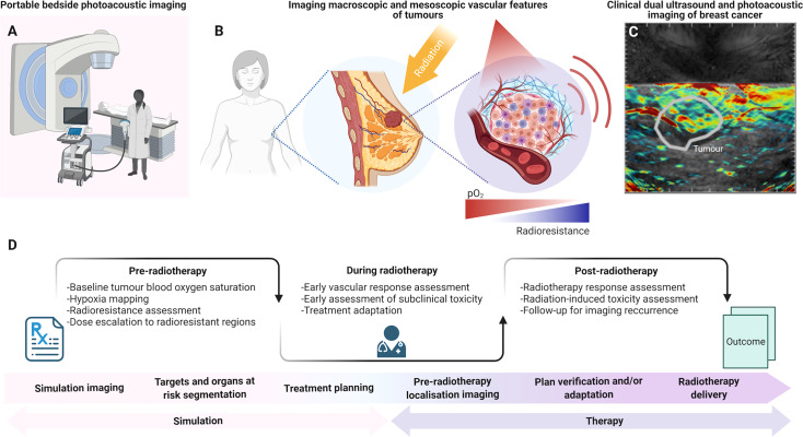
Radiotherapy is recognized globally as a mainstay of treatment in most solid tumors and is essential in both curative and palliative settings. Ionizing radiation is frequently combined with surgery, either preoperatively or postoperatively, and with systemic chemotherapy. Recent advances in imaging have enabled precise targeting of solid lesions yet substantial intratumoral heterogeneity means that treatment planning and monitoring remains a clinical challenge as therapy response can take weeks to manifest on conventional imaging and early indications of progression can be misleading. Photoacoustic imaging (PAI) is an emerging modality for molecular imaging of cancer, enabling non-invasive assessment of endogenous tissue chromophores with optical contrast at unprecedented spatio-temporal resolution. Preclinical studies in mouse models have shown that PAI could be used to assess response to radiotherapy and chemoradiotherapy based on changes in the tumor vascular architecture and blood oxygen saturation, which are closely linked to tumor hypoxia. Given the strong relationship between hypoxia and radio-resistance, PAI assessment of the tumor microenvironment has the potential to be applied longitudinally during radiotherapy to detect resistance at much earlier time-points than currently achieved by size measurements and tailor treatments based on tumor oxygen availability and vascular heterogeneity. Here, we review the current state-of-the-art in PAI in the context of radiotherapy research. Based on these studies, we identify promising applications of PAI in radiation oncology and discuss the future potential and outstanding challenges in the development of translational PAI biomarkers of early response to radiotherapy.
Keywords: image guidance; photoacoustic (optoacoustic) imaging; quantitative imaging biomarker; radiation oncology; radiotherapy; translational research.
Copyright © 2022 Lefebvre, Brown, Hacker, Else, Oraiopoulou, Tomaszewski, Jena and Bohndiek.
Conflict of interest statement
SB has previously received research funding from PreXion Corporation, which (Photoacoustic imaging division) was later acquired by CYBERDYNE Inc. and research support from iThera Medical GmbH, both vendors of photoacoustic imaging equipment. MT would like to disclose that he is currently employed at Merck & Co. The remaining authors declare that the research was conducted in the absence of any commercial or financial relationships that could be construed as a potential conflict of interest.
Figures
Similar articles
-
Noninvasive optoacoustic imaging of breast tumor microvasculature in response to radiotherapy.Front Physiol. 2022 Oct 17;13:1044308. doi: 10.3389/fphys.2022.1044308. eCollection 2022. Front Physiol. 2022. PMID: 36324309 Free PMC article.
-
Photoacoustic Imaging as an Early Biomarker of Radio Therapeutic Efficacy in Head and Neck Cancer.Theranostics. 2018 Mar 6;8(8):2064-2078. doi: 10.7150/thno.21708. eCollection 2018. Theranostics. 2018. PMID: 29721063 Free PMC article.
-
Photoacoustic imaging for investigating tumor hypoxia: a strategic assessment.Theranostics. 2023 May 29;13(10):3346-3367. doi: 10.7150/thno.84253. eCollection 2023. Theranostics. 2023. PMID: 37351178 Free PMC article. Review.
-
Photoacoustic imaging as a highly efficient and precise imaging strategy for the evaluation of brain diseases.Quant Imaging Med Surg. 2021 May;11(5):2169-2186. doi: 10.21037/qims-20-845. Quant Imaging Med Surg. 2021. PMID: 33936997 Free PMC article. Review.
-
Noninvasive monitoring of liver metastasis development via combined multispectral photoacoustic imaging and fluorescence diffuse optical tomography.Int J Biol Sci. 2020 Mar 12;16(9):1616-1628. doi: 10.7150/ijbs.40896. eCollection 2020. Int J Biol Sci. 2020. PMID: 32226306 Free PMC article.
Cited by
-
Performance evaluation of image co-registration methods in photoacoustic mesoscopy of the vasculature.Phys Med Biol. 2024 Sep 25;69(21):215007. doi: 10.1088/1361-6560/ad7fc7. Online ahead of print. Phys Med Biol. 2024. PMID: 39321985 Free PMC article.
-
Niche preclinical and clinical applications of photoacoustic imaging with endogenous contrast.Photoacoustics. 2023 Jul 17;32:100533. doi: 10.1016/j.pacs.2023.100533. eCollection 2023 Aug. Photoacoustics. 2023. PMID: 37636547 Free PMC article. Review.
-
3D Ultrasound-Guided Photoacoustic Imaging to Monitor the Effects of Suboptimal Tyrosine Kinase Inhibitor Therapy in Pancreatic Tumors.Front Oncol. 2022 Jul 7;12:915319. doi: 10.3389/fonc.2022.915319. eCollection 2022. Front Oncol. 2022. PMID: 35875138 Free PMC article.
-
Impact of irradiation conditions on therapy of Lewis lung carcinoma in mice using glucose-ethylenediamine carbon dots.BMC Cancer. 2025 Jan 8;25(1):39. doi: 10.1186/s12885-024-13404-1. BMC Cancer. 2025. PMID: 39780102 Free PMC article.
-
Machine Learning Enabled Photoacoustic Spectroscopy for Noninvasive Assessment of Breast Tumor Progression In Vivo: A Preclinical Study.ACS Sens. 2024 Feb 23;9(2):589-601. doi: 10.1021/acssensors.3c01085. Epub 2024 Jan 30. ACS Sens. 2024. PMID: 38288735 Free PMC article.
References
Grants and funding
LinkOut - more resources
Full Text Sources