

Glycans that regulate Notch signaling in the intestine
- PMID: 35311893
- PMCID: PMC9370068
- DOI: 10.1042/BST20200782
Glycans that regulate Notch signaling in the intestine
Abstract
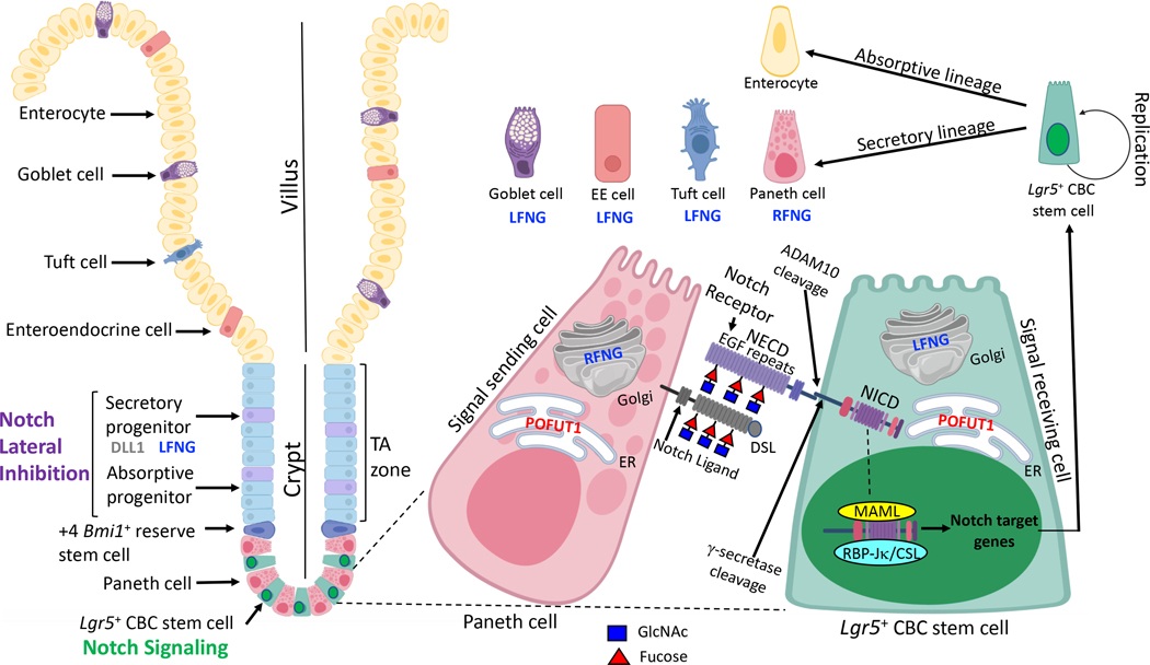
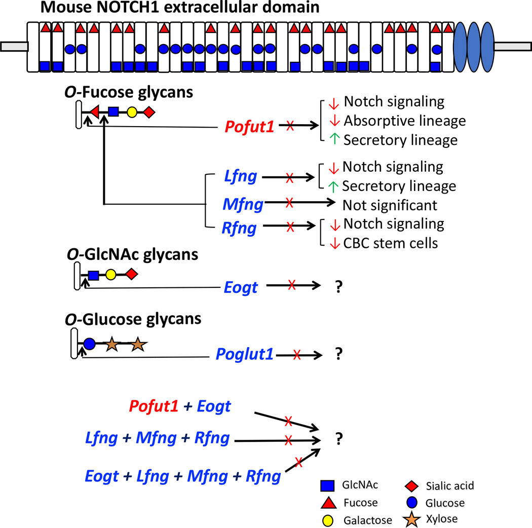
Intestinal homeostasis is key to the maintenance of good health. The small intestine plays important roles in absorption, digestion, hormonal and immune functions. Crypt base columnar (CBC) stem cells residing at the bottom of crypts are nurtured by Paneth cells, and together create the stem cell niche, the foundation of intestinal homeostasis. CBC stem cells replicate to replenish their number, or differentiate into a variety of epithelial cells with specialized functions. Notch signaling is a cell-cell signaling pathway that regulates both the proliferation and differentiation of CBC stem cells. NOTCH1 and NOTCH2 stimulated by canonical Notch ligands DLL1 and DLL4 mediate Notch signaling in the intestine that, in concert with other signaling pathways including the WNT and BMP pathways, determines cell fates. Importantly, interactions between Notch receptors and canonical Notch ligands are regulated by O-glycans linked to Ser/Thr in epidermal growth factor-like (EGF) repeats of the Notch receptor extracellular domain (NECD). The O-glycans attached to NECD are key regulators of the strength of Notch signaling. Imbalances in Notch signaling result in altered cell fate decisions and may lead to cancer in the intestine. In this review, we summarize the impacts of mutations in Notch pathway members on intestinal development and homeostasis, with a focus on the glycosyltransferases that transfer O-glycans to EGF repeats of NOTCH1, NOTCH2, DLL1 and DLL4.
Keywords: Notch signalling pathway; O-glycans; glycosyltransferases; intestinal development; mouse models.
© 2022 The Author(s). Published by Portland Press Limited on behalf of the Biochemical Society.
Figures
Similar articles
-
Regulation of Notch signaling during T- and B-cell development by O-fucose glycans.Immunol Rev. 2009 Jul;230(1):201-15. doi: 10.1111/j.1600-065X.2009.00791.x. Immunol Rev. 2009. PMID: 19594638 Review.
-
Regulation of myeloid and lymphoid cell development by O-glycans on Notch.Front Mol Biosci. 2022 Nov 4;9:979724. doi: 10.3389/fmolb.2022.979724. eCollection 2022. Front Mol Biosci. 2022. PMID: 36406268 Free PMC article. Review.
-
EOGT enables residual Notch signaling in mouse intestinal cells lacking POFUT1.Sci Rep. 2023 Oct 14;13(1):17473. doi: 10.1038/s41598-023-44509-5. Sci Rep. 2023. PMID: 37838775 Free PMC article.
-
Multiple roles for O-glycans in Notch signalling.FEBS Lett. 2018 Dec;592(23):3819-3834. doi: 10.1002/1873-3468.13251. Epub 2018 Nov 28. FEBS Lett. 2018. PMID: 30207383 Free PMC article. Review.
-
Xylosyl Extension of O-Glucose Glycans on the Extracellular Domain of NOTCH1 and NOTCH2 Regulates Notch Cell Surface Trafficking.Cells. 2020 May 14;9(5):1220. doi: 10.3390/cells9051220. Cells. 2020. PMID: 32423029 Free PMC article.
Cited by
-
Pathways regulating intestinal stem cells and potential therapeutic targets for radiation enteropathy.Mol Biomed. 2024 Oct 10;5(1):46. doi: 10.1186/s43556-024-00211-0. Mol Biomed. 2024. PMID: 39388072 Free PMC article. Review.
-
RNA-Seq data provide new insights into the molecular regulation of breast muscle glycogen reserves, a key factor in muscle function and meat quality in chickens.Poult Sci. 2025 Jun;104(6):105136. doi: 10.1016/j.psj.2025.105136. Epub 2025 Apr 4. Poult Sci. 2025. PMID: 40215882 Free PMC article.
-
Butyrate alleviates food allergy by improving intestinal barrier integrity through suppressing oxidative stress-mediated Notch signaling.Imeta. 2025 Apr 3;4(3):e70024. doi: 10.1002/imt2.70024. eCollection 2025 Jun. Imeta. 2025. PMID: 40469511 Free PMC article.
-
Molecular regulation after mucosal injury and regeneration in ulcerative colitis.Front Mol Biosci. 2022 Oct 13;9:996057. doi: 10.3389/fmolb.2022.996057. eCollection 2022. Front Mol Biosci. 2022. PMID: 36310594 Free PMC article. Review.
-
Mechanism of Notch Signaling Pathway in Malignant Progression of Glioblastoma and Targeted Therapy.Biomolecules. 2024 Apr 15;14(4):480. doi: 10.3390/biom14040480. Biomolecules. 2024. PMID: 38672496 Free PMC article. Review.
References
-
- Cheng H, Leblond CP. Origin, differentiation and renewal of the four main epithelial cell types in the mouse small intestine. V. Unitarian Theory of the origin of the four epithelial cell types. Am J Anat. 1974;141(4):537–61. - PubMed
-
- Hosoyamada Y, Sakai T. Structural and mechanical architecture of the intestinal villi and crypts in the rat intestine: integrative reevaluation from ultrastructural analysis. Anatomy and embryology. 2005;210(1):1–12. - PubMed
-
- Barker N Adult intestinal stem cells: critical drivers of epithelial homeostasis and regeneration. Nat Rev Mol Cell Biol. 2014;15(1):19–33. - PubMed
-
- Gehart H, Clevers H. Tales from the crypt: new insights into intestinal stem cells. Nature Reviews Gastroenterology & Hepatology. 2019;16(1):19–34. - PubMed
Publication types
MeSH terms
Substances
Grants and funding
LinkOut - more resources
Full Text Sources
Miscellaneous