

Modeling the catarrhal stage of Bordetella pertussis upper respiratory tract infections in mice
- PMID: 35311902
- PMCID: PMC9092653
- DOI: 10.1242/dmm.049266
Modeling the catarrhal stage of Bordetella pertussis upper respiratory tract infections in mice
Abstract
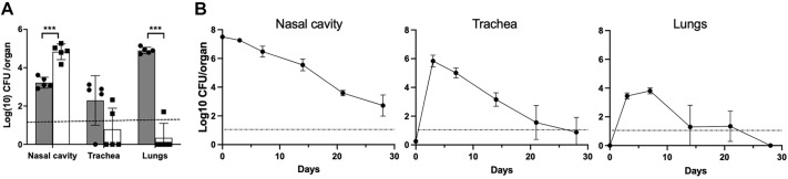
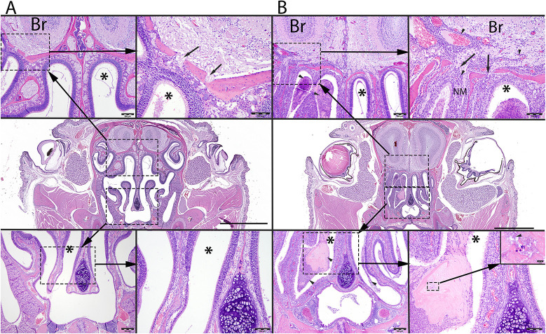
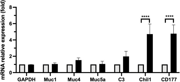
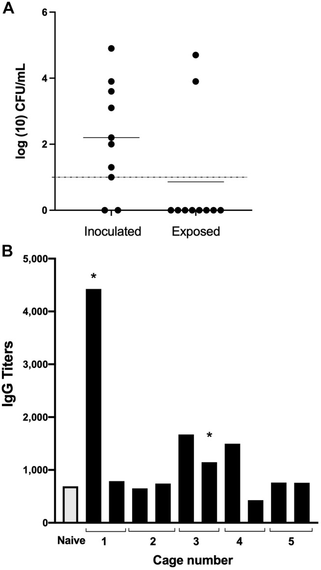
Pertussis (whooping cough) is a highly transmissible human respiratory disease caused by Bordetella pertussis, a human-restricted pathogen. Animal models generally involve pneumonic infections induced by depositing large numbers of bacteria in the lungs of mice. These models have informed us about the molecular pathogenesis of pertussis and guided development of vaccines that successfully protect against severe disease. However, they bypass the catarrhal stage of the disease, when bacteria first colonize and initially grow in the upper respiratory tract. This is a critical and highly transmissible stage of the infection that current vaccines do not prevent. Here, we demonstrate a model system in which B. pertussis robustly and persistently infects the nasopharynx of TLR4-deficient mice, inducing localized inflammation, neutrophil recruitment and mucus production as well as persistent shedding and occasional transmission to cage mates. This novel experimental system will allow the study of the contributions of bacterial factors to colonization of and shedding from the nasopharynx, as occurs during the catarrhal stage of pertussis, and interventions that might better control the ongoing circulation of pertussis.
Keywords: Bordetella pertussis; Catarrhal stage; Mouse; Shedding; TLR4 receptor.
© 2022. Published by The Company of Biologists Ltd.
Conflict of interest statement
Competing interests The authors declare no competing or financial interests.
Figures
Similar articles
-
Modeling Immune Evasion and Vaccine Limitations by Targeted Nasopharyngeal Bordetella pertussis Inoculation in Mice.Emerg Infect Dis. 2021 Aug;27(8):2107-2116. doi: 10.3201/eid2708.203566. Emerg Infect Dis. 2021. PMID: 34286682 Free PMC article.
-
Bordetella pertussis filamentous hemagglutinin: evaluation as a protective antigen and colonization factor in a mouse respiratory infection model.Infect Immun. 1990 Jan;58(1):7-16. doi: 10.1128/iai.58.1.7-16.1990. Infect Immun. 1990. PMID: 2294058 Free PMC article.
-
Protection against intranasal infection of mice with Bordetella pertussis.Dev Biol Stand. 1985;61:165-72. Dev Biol Stand. 1985. PMID: 2872102
-
Bordetella pertussis and pertactin-deficient clinical isolates: lessons for pertussis vaccines.Expert Rev Vaccines. 2014 Sep;13(9):1135-46. doi: 10.1586/14760584.2014.932254. Epub 2014 Jun 23. Expert Rev Vaccines. 2014. PMID: 24953157 Review.
-
The Role of Mucosal Immunity in Pertussis.Front Immunol. 2019 Jan 14;9:3068. doi: 10.3389/fimmu.2018.03068. eCollection 2018. Front Immunol. 2019. PMID: 30692990 Free PMC article. Review.
Cited by
-
Bordetella pertussis infection activates the type I interferon signaling pathway to exacerbate respiratory tract inflammatory response.Front Immunol. 2025 Mar 7;16:1521970. doi: 10.3389/fimmu.2025.1521970. eCollection 2025. Front Immunol. 2025. PMID: 40124358 Free PMC article.
-
Bordetella spp. block eosinophil recruitment to suppress the generation of early mucosal protection.Cell Rep. 2023 Nov 28;42(11):113294. doi: 10.1016/j.celrep.2023.113294. Epub 2023 Oct 25. Cell Rep. 2023. PMID: 37883230 Free PMC article.
-
The Fim and FhaB adhesins play a crucial role in nasal cavity infection and Bordetella pertussis transmission in a novel mouse catarrhal infection model.PLoS Pathog. 2022 Apr 8;18(4):e1010402. doi: 10.1371/journal.ppat.1010402. eCollection 2022 Apr. PLoS Pathog. 2022. PMID: 35395059 Free PMC article.
-
Generating enhanced mucosal immunity against Bordetella pertussis: current challenges and new directions.Front Immunol. 2023 Feb 21;14:1126107. doi: 10.3389/fimmu.2023.1126107. eCollection 2023. Front Immunol. 2023. PMID: 36895562 Free PMC article.
-
Intranasal challenge with B. pertussis leads to more severe disease manifestations in mice than aerosol challenge.PLoS One. 2023 Nov 2;18(11):e0286925. doi: 10.1371/journal.pone.0286925. eCollection 2023. PLoS One. 2023. PMID: 37917623 Free PMC article.
References
-
- Allen, A. C., Wilk, M. M., Misiak, A., Borkner, L., Murphy, D. and Mills, K. H. G. (2018). Sustained protective immunity against Bordetella pertussis nasal colonization by intranasal immunization with a vaccine-adjuvant combination that induces IL-17-secreting TRM cells. Mucosal Immunol. 6, 1763-1776. 10.1038/s41385-018-0080-x - DOI - PubMed
-
- Bayat, B., Werth, S., Sachs, U. J., Newman, D. K., Newman, P. J. and Santoso, S. (2010). Neutrophil transmigration mediated by the neutrophil-specific antigen CD177 is influenced by the endothelial S536N dimorphism of platelet endothelial cell adhesion molecule-1. J. Immunol. 184, 3889-3896. 10.4049/jimmunol.0903136 - DOI - PMC - PubMed
-
- Cherry, J. D. (2015). The history of pertussis (whooping cough); 1906-2015: facts, myths, and misconceptions. Curr. Epidemiol. Rep. 2, 120-130. 10.1007/s40471-015-0041-9 - DOI
Publication types
MeSH terms
Substances
Grants and funding
LinkOut - more resources
Full Text Sources
Medical
