Effect of Tranexamic Acid Administration on Remote Cerebral Ischemic Lesions in Acute Spontaneous Intracerebral Hemorrhage: A Substudy of a Randomized Clinical Trial
- PMID: 35311937
- PMCID: PMC8938900
- DOI: 10.1001/jamaneurol.2022.0217
Effect of Tranexamic Acid Administration on Remote Cerebral Ischemic Lesions in Acute Spontaneous Intracerebral Hemorrhage: A Substudy of a Randomized Clinical Trial
Abstract
Importance: Hyperintense foci on diffusion-weighted imaging (DWI) that are spatially remote from the acute hematoma occur in 20% of people with acute spontaneous intracerebral hemorrhage (ICH). Tranexamic acid, a hemostatic agent that is under investigation for treating acute ICH, might increase DWI hyperintense lesions (DWIHLs).
Objective: To establish whether tranexamic acid compared with placebo increased the prevalence or number of remote cerebral DWIHLs within 2 weeks of ICH onset.
Design, setting, and participants: This prospective nested magnetic resonance imaging (MRI) substudy of a randomized clinical trial (RCT) recruited participants from the multicenter, double-blind, placebo-controlled, phase 3 RCT (Tranexamic Acid for Hyperacute Primary Intracerebral Hemorrhage [TICH-2]) from July 1, 2015, to September 30, 2017, and conducted follow-up to 90 days after participants were randomized to either the tranexamic acid or placebo group. Participants had acute spontaneous ICH and included TICH-2 participants who provided consent to undergo additional MRI scans for the MRI substudy and those who had clinical MRI data that were compatible with the brain MRI protocol of the substudy. Data analyses were performed on an intention-to-treat basis on January 20, 2020.
Interventions: The tranexamic acid group received 1 g in 100-mL intravenous bolus loading dose, followed by 1 g in 250-mL infusion within 8 hours of ICH onset. The placebo group received 0.9% saline within 8 hours of ICH onset. Brain MRI scans, including DWI, were performed within 2 weeks.
Main outcomes and measures: Prevalence and number of remote DWIHLs were compared between the treatment groups using binary logistic regression adjusted for baseline covariates.
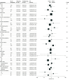
Results: A total of 219 participants (mean [SD] age, 65.1 [13.8] years; 126 men [57.5%]) who had brain MRI data were included. Of these participants, 96 (43.8%) were randomized to receive tranexamic acid and 123 (56.2%) were randomized to receive placebo. No baseline differences in demographic characteristics and clinical or imaging features were found between the groups. There was no increase for the tranexamic acid group compared with the placebo group in DWIHL prevalence (20 of 96 [20.8%] vs 28 of 123 [22.8%]; odds ratio [OR], 0.71; 95% CI, 0.33-1.53; P = .39) or mean (SD) number of DWIHLs (1.75 [1.45] vs 1.81 [1.71]; mean difference [MD], -0.08; 95% CI, -0.36 to 0.20; P = .59). In an exploratory analysis, participants who were randomized within 3 hours of ICH onset or those with chronic infarcts appeared less likely to have DWIHLs if they received tranexamic acid. Participants with probable cerebral amyloid angiopathy appeared more likely to have DWIHLs if they received tranexamic acid.
Conclusions and relevance: This substudy of an RCT found no evidence of increased prevalence or number of remote DWIHLs after tranexamic acid treatment in acute ICH. These findings provide reassurance for ongoing and future trials that tranexamic acid for acute ICH is unlikely to induce cerebral ischemic events.
Trial registration: isrctn.org Identifier: ISRCTN93732214.
Conflict of interest statement
Figures


Comment in
-
Tranexamic Acid and Diffusion-Weighted Imaging Lesions After Intracerebral Hemorrhage.JAMA Neurol. 2022 May 1;79(5):447-449. doi: 10.1001/jamaneurol.2022.0228. JAMA Neurol. 2022. PMID: 35311924 No abstract available.

