. 2022 Apr 1;149(7):dev198341.
doi: 10.1242/dev.198341. Epub 2022 Apr 14.

Molecular contribution to embryonic aneuploidy and karyotypic complexity in initial cleavage divisions of mammalian development

Affiliations

Molecular contribution to embryonic aneuploidy and karyotypic complexity in initial cleavage divisions of mammalian development

Kelsey E Brooks et al. Development. .

Abstract

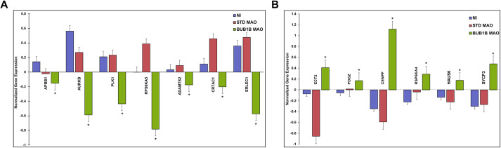
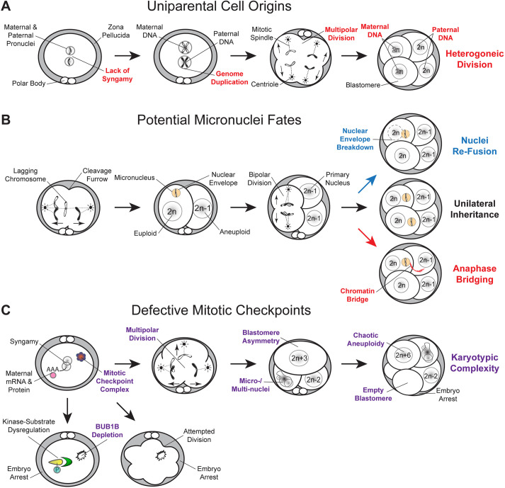
Embryonic aneuploidy is highly complex, often leading to developmental arrest, implantation failure or spontaneous miscarriage in both natural and assisted reproduction. Despite our knowledge of mitotic mis-segregation in somatic cells, the molecular pathways regulating chromosome fidelity during the error-prone cleavage-stage of mammalian embryogenesis remain largely undefined. Using bovine embryos and live-cell fluorescent imaging, we observed frequent micro-/multi-nucleation of mis-segregated chromosomes in initial mitotic divisions that underwent unilateral inheritance, re-fused with the primary nucleus or formed a chromatin bridge with neighboring cells. A correlation between a lack of syngamy, multipolar divisions and asymmetric genome partitioning was also revealed, and single-cell DNA-seq showed propagation of primarily non-reciprocal mitotic errors. Depletion of the mitotic checkpoint protein BUB1B (also known as BUBR1) resulted in similarly abnormal nuclear structures and cell divisions, as well as chaotic aneuploidy and dysregulation of the kinase-substrate network that mediates mitotic progression, all before zygotic genome activation. This demonstrates that embryonic micronuclei sustain multiple fates, provides an explanation for blastomeres with uniparental origins, and substantiates defective checkpoints and likely other maternally derived factors as major contributors to the karyotypic complexity afflicting mammalian preimplantation development.

Keywords: Aneuploidy; BUB1B; BUBR1; Cytokinesis; Embryo; Micronuclei; Mitosis.


Conflict of interest statement

Competing interests The authors declare no competing or financial interests.

Figures

Fig. 1.
Fig. 1.
Investigating the dynamics of mitotic chromosome segregation and MCC fidelity in bovine embryos. (A) In vitro-produced bovine oocytes underwent IVF and the resulting zygotes were non-invasively monitored by time-lapse image analysis until collection for immunostaining of nuclear structure (n=53). Another subset of zygotes was microinjected with fluorescently labeled modified mRNAs and chromosome segregation visualized during the first three mitotic divisions in real-time by live-cell confocal microscopy (n=90). Embryos were disassembled into single blastomeres at the 2- to 12-cell stage for scDNA-seq and CNV analysis to determine the precise frequency of aneuploidy at the early cleavage stages (n=38). Other zygotes were microinjected with non-overlapping morpholinos targeting the mitotic checkpoint protein BUB1B and/or modified BUB1B mRNA to test the effect and specificity of MCC inhibition on chromosome segregation, division dynamics and preimplantation development (n=430). Gene expression profiling was also conducted on a subset of MCC-deficient zygotes versus controls by quantitative RT-PCR to identify changes in gene abundance and molecular pathways associated with BUB1B knockdown (n=15). (B,C) Immunostaining of zygotes (B) and cleavage-stage embryos (C) with LMNB1 (green) using DAPI (blue) to visualize DNA revealed several micro- and multi-nuclei (white arrowheads). (D,E) Blastocysts also immunostained for the trophoblast marker CDX2 (red) showed that micronuclei (arrowheads) are often present in the TE (E), but can also be retained within the ICM of the embryo. In D, the micronucleus is present in the TE; in E, micronuclei are present in both the TE (top left) and the ICM (bottom right). Scale bars: 10 µm.
Fig. 2.
Fig. 2.
Live-cell fluorescent imaging reveals micronuclei fate and uniparental genome distribution to daughter cells. Bovine zygotes were microinjected with fluorescently labeled modified mRNAs (mCitrine or mCherry) to visualize DNA (Histone H2B) or nuclear structure (LMNB1) and distinguish blastomeres (F-actin) by live-cell confocal microscopy during the first three mitotic divisions (n=90). (A) A Venn Pie that shows the percentage of embryos that did not complete cytokinesis (gray), exhibited normal bipolar divisions (orange) or underwent multipolar divisions at the zygote or two-cell stage (pink). The percentage of embryos with micro- and/or multi-nuclei (MN; yellow) associated with each type of division is also shown. Micronuclei fate is represented as those that formed a chromatin bridge (dark blue), exhibited unilateral inheritance (medium blue) or re-fused with the primary nucleus (light blue). Most embryos underwent bipolar divisions and were more likely to contain micronuclei than multipolar embryos. (B,C) Anaphase lagging of chromosomes (white arrowheads) was detected in certain embryos at the zygote (B) or two-cell (C) stage before micronuclei formation. (D-F) An examination of micronuclei fate demonstrated that relatively equal proportions persist and undergo unilateral inheritance (D) or fuse back with the primary nucleus (E), with a small number exhibiting what appeared to be a chromatin bridge between blastomeres following micronuclei formation (F) (white arrowheads). (G,H) The majority of multipolar embryos (white solid arrows) bypassed pronuclear fusion (syngamy) before the abnormal division (G) and often produced blastomeres with asymmetric genome partitioning and/or no apparent nuclear structure (H, white dashed arrows). Numbers in the top left corner indicate the time since the start of imaging; the auto-labeling of each embryo used for identification and/or tracking purposes has been masked for clarity where necessary.
Fig. 3.
Fig. 3.
Comprehensive assessment of chromosomal abnormalities in early cleavage-stage embryos by scDNA-seq. (A) Whole-chromosome and sub-chromosomal CNV was evaluated in bovine embryos from the 2- to 12-cell stage (n=38). Stacked bars represent all blastomeres (n=133) classified as euploid (green), aneuploid (blue), segmental aneuploid (purple), chaotic aneuploid (yellow), empty (gray) or failing to undergo WGA (white). (B) Pie chart showing the overall chromosome status of the embryos. (C) Number of whole or segmental chromosome losses and/or gains affecting each chromosome. There is frequent mis-segregation of the X-chromosome and DNA breakage in chromosomes 5, 7, 11 and 29. (D) The percentage of aneuploid embryos with each type of chromosomal error. (E) CNV plots of blastomeres from two different embryos with chaotic aneuploidy showing up to six copies of some chromosomes (top; black solid arrow) and a complete loss of other chromosomes (bottom; black dashed arrow). (F) Blastomeres from a two-cell embryo with meiotic errors (chromosomes 7, 8 and 9) propagated during the first cleavage division that also experienced mitotic mis-segregation of different chromosomes (chromosomes 19 and 21) that were reciprocal.
Fig. 4.
Fig. 4.
BUB1B knockdown induces multipolar divisions, chaotic aneuploidy and developmental arrest. (A,B) Darkfield time-lapse imaging frames depicting the various embryo phenotypes (red arrows), including attempted division, multipolar division and blastomere asymmetry observed following BUB1B MAO 1 (A) or BUB1B MAO 2 (B) microinjection in bovine zygotes. (C) Representative stereomicroscope images of embryos and blastocysts from the Std Control MAO, BUB1B MAO 1, and BUB1B MAO 1 and BUB1B modified mRNA treatment groups. (D) Bar graph of the percentage of embryos that reached the blastocyst stage in non-injected (n=180), Std Control MAO- (n=81), BUB1B MAO 1- (n=48), BUB1B MAO 2- (n=36), or BUB1B MAO 1- and BUB1B modified mRNA-injected zygotes (n=85). Although no blastocysts were obtained following BUB1B MAO 1 or 2 treatment, the co-injection of BUB1 MAO 1 and BUB1B modified mRNA was able to almost fully rescue the phenotype and restore blastocyst formation rates to that observed in controls. (E) Confocal images of LMNB1 (green) immunostaining in BUB1B MAO 1- or 2-treated embryos stained with DAPI (blue). Abnormal nuclear morphology and the presence of both micro- and multi-nuclei were detected (white arrowheads) in embryos at the zygote stage (top row) and cleavage stage that exhibited abnormal cell divisions (bottom row). DNA without a nuclear envelope (white arrows) and a blastomere that completely lacked nuclear material are present in the two-cell embryo located in the lower-left image. Scale bars: 10 µm. (F) CNV plots of blastomeres from different cleavage-stage embryos disassembled into single cells following BUB1B 1 MAO injection. Although some euploid blastomeres were detected in BUB1B-injected embryos (upper left plot), most exhibited chaotic aneuploidy with multiple whole and sub-chromosomal losses and gains.
Fig. 5.
Fig. 5.
BUB1B deficiency in zygotes primarily impacts the expression of other protein kinases and kinase substrates. The relative abundance of several mitotic, cell cycle, developmentally regulated and cell survival genes was assessed via microfluidic quantitative RT-PCR (RT-qPCR) in non-injected (NI; n=5), Std Control MAO-treated (n=5) and BUB1B MAO 1-treated (n=5) individual zygotes using gene-specific primers. (A) The genes that were significantly downregulated (*P≤0.05) in BUB1B MAO-injected embryos compared with the NI and Std Control MAO are shown in the bar graph (data are mean±s.e.m.). (B) A bar graph of the genes that were significantly upregulated (*P≤0.05) in BUB1B MAO-injected embryos relative to the controls (data are mean±s.e.m.). Mean CNRQ values for each gene were compared across embryo groups using the Mann–Whitney U-test. The full list of the 96 genes and primer sequences assessed by RT-qPCR is available in Fig. S4 and Table S2, respectively.
Fig. 6.
Fig. 6.
Summary of the major conclusions from the imaging, scDNA-seq and gene knockdown studies. (A) Simplified model of how the lack of maternal and paternal pronuclear fusion (syngamy) at the zygote stage, followed by genome duplication and multipolar divisions, could contribute to blastomeres with uniparental origins or to those that contain only maternal or paternal DNA. (B) Live-cell imaging also revealed the formation of anaphase lagging chromosomes likely from merotelic attachments before or during the first mitotic division. The chromosome(s) become encapsulated in nuclear envelope to form a micronucleus and the embryo continues to divide normally. In these subsequent bipolar divisions, most micronuclei either fuse back with the primary nucleus upon nuclear envelope breakdown or persist and undergo unilateral inheritance; however, some micronuclei form a chromatin bridge with the nucleus of another blastomere during anaphase. (C) The depletion of BUB1B in zygotes resulted in no division or attempted division and in embryo arrest, while multipolar divisions, blastomere asymmetry and micro-/multi-nuclei were observed in MCC-deficient embryos that completed the first cytokinesis. These abnormal divisions also produced daughter cells with chaotic aneuploidy and/or empty blastomeres with no nuclear structure that induced embryo arrest, which suggested that the lack of MCC permits the karyotypic complexity detected at the early cleavage-stages of preimplantation development.

References

    1. Alper, M. M., Brinsden, P., Fischer, R. and Wikland, M. (2001). To blastocyst or not to blastocyst? That is the question. Hum. Reprod. 16, 617-619. 10.1093/humrep/16.4.617 - DOI - PubMed
    1. Baker, D. J., Jeganathan, K. B., Cameron, J. D., Thompson, M., Juneja, S., Kopecka, A., Kumar, R., Jenkins, R. B., de Groen, P. C., Roche, P.et al. (2004). BubR1 insufficiency causes early onset of aging-associated phenotypes and infertility in mice. Nat. Genet. 36, 744-749. 10.1038/ng1382 - DOI - PubMed
    1. Baker, D. J., Dawlaty, M. M., Wijshake, T., Jeganathan, K. B., Malureanu, L., van Ree, J. H., Crespo-Diaz, R., Reyes, S., Seaburg, L., Shapiro, V.et al. (2013). Increased expression of BubR1 protects against aneuploidy and cancer and extends healthy lifespan. Nat. Cell Biol. 15, 96-102. 10.1038/ncb2643 - DOI - PMC - PubMed
    1. Baltaci, V., Satiroglu, H., Kabukçu, C., Ünsal, E., Aydinuraz, B., Üner, O., Aktas, Y., Çetinkaya, E., Turhan, F. and Aktan, A. (2006). Relationship between embryo quality and aneuploidies. Reprod. Biomed. Online 12, 77-82. 10.1016/S1472-6483(10)60984-4 - DOI - PubMed
    1. Bibi, N., Parveen, Z. and Rashid, S. (2013). Identification of potential Plk1 targets in a cell-cycle specific proteome through structural dynamics of kinase and Polo box-mediated interactions. PLoS ONE 8, e70843. 10.1371/journal.pone.0070843 - DOI - PMC - PubMed

Publication types