. 2022 Apr 18;35(4):636-650.
doi: 10.1021/acs.chemrestox.1c00420. Epub 2022 Mar 21.

Nitrogen Mustard Alkylates and Cross-Links p53 in Human Keratinocytes

Affiliations

Nitrogen Mustard Alkylates and Cross-Links p53 in Human Keratinocytes

Yi-Hua Jan et al. Chem Res Toxicol. .

Abstract

Cytotoxic blistering agents such as sulfur mustard and nitrogen mustard (HN2) were synthesized for chemical warfare. Toxicity is due to reactive chloroethyl side chains that modify and damage cellular macromolecules including DNA and proteins. In response to DNA damage, cells initiate a DNA damage response directed at the recruitment and activation of repair-related proteins. A central mediator of the DNA damage response is p53, a protein that plays a critical role in regulating DNA repair. We found that HN2 causes cytosolic and nuclear accumulation of p53 in HaCaT keratinocytes; HN2 also induced post-translational modifications on p53 including S15 phosphorylation and K382 acetylation, which enhance p53 stability, promote DNA repair, and mediate cellular metabolic responses to stress. HN2 also cross-linked p53, forming dimers and high-molecular-weight protein complexes in the cells. Cross-linked multimers were also modified by K48-linked ubiquitination indicating that they are targets for proteasome degradation. HN2-induced modifications transiently suppressed the transcriptional activity of p53. Using recombinant human p53, HN2 alkylation was found to be concentration- and redox status-dependent. Dithiothreitol-reduced protein was more efficiently cross-linked indicating that p53 cysteine residues play a key role in protein modification. LC-MS/MS analysis revealed that HN2 directly alkylated p53 at C124, C135, C141, C176, C182, C275, C277, H115, H178, K132, and K139, forming both monoadducts and cross-links. The formation of intermolecular complexes was a consequence of HN2 cross-linked cysteine residues between two molecules of p53. Together, these data demonstrate that p53 is a molecular target for mustard vesicants. Modification of p53 likely mediates cellular responses to HN2 including DNA repair and cell survival contributing to vesicant-induced cytotoxicity.


Conflict of interest statement

The authors declare no competing financial interest.

Figures
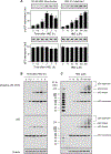
Figure 1.
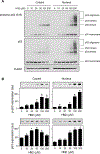
Figure 1.. HN2 cross-links p53 proteins in HaCaT cells.
Cells were treated with increasing concentrations of HN2 or vehicle control in serum-free medium. At the indicated times, cells were harvested, and total cellular lysates were prepared; protein expression was analyzed by (A) slot blots or (B, C) by SDS-polyacrylamide gel electrophoresis in reducing and denaturing gels followed by Western blotting. β-Actin was used as an example of a protein loading control. Representative Western blots and slot blots from one of three separate experiments are shown. Data are the mean ± SE (n = 3). *Significantly different (p < 0.05) from vehicle-treated controls. (B) Time-course of changes in p53 proteins in response to 50 μM HN2. (C) Effects of increasing concentrations of HN2 on the expression of p53 proteins. Protein expression was analyzed 2 h after HN2 treatment.
Figure 2.
Figure 2.. Cellular cross-linking of p53 by bifunctional alkylating agents.
(A) HaCaT cells were incubated with 200 μM chlorambucil (CHL), melphalan (MEL), or cisplatin (CDDP) in serum-free medium. At the indicated times (0–6 h), cells were harvested, and cell lysates were prepared; p53 protein expression was analyzed by Western blotting. β-Actin was used as a protein loading control. Little to no cytotoxicity was observed after a 6 h exposure of cells to these agents (200 μM) as measured by the PrestoBlue assay. (B) Effects of HN2 on p53 cross-linking in cells with wild-type p53 and mutant p53. Cells were treated without and with HN2 (200 μM). After 2 h, lysates were prepared and analyzed for p53 cross-linking by Western blotting. Cell lines used include HaCaT (mutant p53 with H179Y and R282W mutations), HEK-293 (wild-type p53), CX-1 and A431 (mutant p53 with R273H mutation), and MLE-15 (wild-type p53). Data show representative Western blots from one of three separate experiments.
Figure 3.
Figure 3.. Effects of HN2 on cytoplasmic and nuclear localization of p53 in HaCaT cells.
Cells were treated with increasing concentrations of HN2 (10–200 μM) or vehicle control in serum-free medium. After 2 h, cytosolic and nuclear fractions were prepared, and p53 and phospho p53 expression was analyzed by Western blotting and slot blotting. (A) HN2 cross-links p53 proteins in both cytosolic and nuclear fractions of HaCaT cells. Representative Western blots from three separate experiments are shown. β-Actin was used as a protein loading control. (B) Relative phospho p53 (S15) and p53 protein expression in HaCaT cells after HN2 treatment analyzed by slot blotting. Representative slot blots from three separate experiments are shown. Data are the mean ± SE (n = 3). *Significantly different (p < 0.05) from vehicle-treated controls.
Figure 4.
Figure 4.. Effects of HN2 on transcriptional activity and post-translational modification of p53 in HaCaT cells.
Cells were treated with increasing concentrations of HN2 (10–200 μM) or vehicle control. After 2 h, whole cell lysates or nuclear fractions of cells were prepared. (A) Nuclear fractions were assayed for p53 transcriptional activity as described in the Materials and Methods section. Data are the mean ± SE (n = 3). *Significantly different (p < 0.05) from vehicle-treated controls. Cell lysates were analyzed by Western blotting for acetylation of p53 at K382 (B) and ubiquitination as indicated (C). β-Actin was used as an example of a loading control. (D) Ubiquitination assay of p53. Whole cell lysates were subjected to immunoprecipitation with a monoclonal antibody against p53 conjugated to agarose beads. K48-linked polyubiquitinated p53 and p53 in the immunoprecipitates were determined by Western blotting.
Figure 5.
Figure 5.. Effects of HN2 on recombinant human p53.
Recombinant wild-type human p53 (100 nM) was incubated without and with DTT (10 mM) at 37 °C. After 20 min, proteins were purified using Chroma Spin TE-10 columns to remove DTT and then incubated with increasing concentrations of HN2 (2–200 μM) or vehicle control in potassium phosphate buffer (100 mM, pH 7.4) at room temperature. After an additional 60 min, samples were analyzed by SDS-polyacrylamide gel electrophoresis under reducing and denaturing conditions followed by Western blotting.
Figure 6.
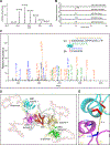
Figure 6.. Mass spectrum of HN2 cross-linked peptides at m/z 494.8439.
(A) Mass spectrum of the quintuply charged cross-linked peptides with monoisotopic m/z 494.8439. (B) Extracted-ion chromatogram of the parent ion in different p53 protein bands. (C) Higher-energy collisional dissociation (HCD) fragmentation of the [M + 5H]5+ precursor ion at m/z 494.8439. The sequences of cross-linked peptides were identified as CSDSDGLAPPQHLIR (residues 182–196 on p53; α peptide) and CPHHER (residues 176–181 on p53; β peptide). Identified fragments are indicated with α or β to denote the peptide from which they originated, together with their charge states. This cross-link involves residues C182 and C176 from α and β peptide, respectively. The inset shows the cross-linked fragments with the identified b and y ions labeled. (D) Stereo view of the structure of full-length wild-type human p53 tetramer. The molecular model was based on the SAXS model of human p53 in the absence of DNA. The subdomains of the DNA binding domain (DBD) are shown in cyan, green, magenta, and blue, respectively, with ribbon representation. Oligomerization domains (ODs) are shown in red. (E) Close-up of the interface of two p53 DBD subunits. Distances (Å) between thiol groups of C176 and C182 are labeled.
Figure 7.
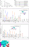
Figure 7.. Mass spectrum of HN2 cross-linked peptides at m/z 532.7640.
(A) Mass spectrum of the quadruply charged cross-linked peptides with monoisotopic m/z 532.7640. (B) Extracted-ion chromatogram of the precursor ion at m/z 532.7640 in different p53 protein bands. (C, D) HCD fragmentation of the [M + 4H]4+ m/z 532.7640 precursor ion. The sequences of cross-linked peptides were identified as SVTCTYSPALNK (residues 121–132 on p53; α peptide) and VCACPGR (residues 274–280 on p53; β peptide) with one HN2 cross-link and one carbamidomethylation (cam) on cysteine. Identified fragments are indicated with α or β to denote the peptide from which they originated, together with their charge states. This cross-link involves residues C124 and C275 (C) or C124 and C277 (D) from the α and β peptide, respectively. The inset shows the cross-linked fragments with the identified b and y ions labeled. (E) Cross-linking sites on the SAXS model of human p53. Cross-linked and/or carbamidomethylated residues C124, C275, and C277 (colored in red) are located at the surface of the p53 structure. The distances (Å) between thiol groups of cysteine residues are labeled.
Figure 8.
Figure 8.. Molecular model of human p53 core DNA binding domain.
(A) Location of cysteine residues in the p53 DNA binding domain (top) and relative distances between the thiol groups of cysteine residues in the p53 model (bottom). Cysteine residues from one subunit are shown in a stick representation in red. (B) Surface of the p53 core domain (top) and solvent accessibility surface of cysteine residues in the p53 model (bottom). Solvent-exposed cysteine residues are shown in red.

Similar articles

Cited by

References

    1. Dacre JC; Goldman M Toxicology and pharmacology of the chemical warfare agent sulfur mustard. Pharmacol. Rev. 1996, 48 (2), 289–326. - PubMed
    1. Singh RK; Kumar S; Prasad DN; Bhardwaj TR Therapeutic journery of nitrogen mustard as alkylating anticancer agents: Historic to future perspectives. Eur. J. Med. Chem. 2018, 151, 401–433. - PubMed
    1. Balali-Mood M; Hefazi M The pharmacology, toxicology, and medical treatment of sulphur mustard poisoning. Fundam Clin Pharmacol 2005, 19 (3), 297–315. DOI: 10.1111/j.1472-8206.2005.00325.x. - DOI - PubMed
    1. Jan YH; Heck DE; Laskin DL; Laskin JD DNA damage signaling in the cellular responses to mustard vesicants. Toxicol. Lett. 2020, 326, 78–82. - PMC - PubMed
    1. Shakarjian MP; Heck DE; Gray JP; Sinko PJ; Gordon MK; Casillas RP; Heindel ND; Gerecke DR; Laskin DL; Laskin JD Mechanisms mediating the vesicant actions of sulfur mustard after cutaneous exposure. Toxicol. Sci. 2010, 114 (1), 5–19. - PMC - PubMed

Publication types