. 2022 Mar 29;119(13):e2119509119.
doi: 10.1073/pnas.2119509119. Epub 2022 Mar 21.

Functional enzyme-polymer complexes

Affiliations

Functional enzyme-polymer complexes

Curt Waltmann et al. Proc Natl Acad Sci U S A. .

Abstract

SignificanceThe use of biological enzyme catalysts could have huge ramifications for chemical industries. However, these enzymes are often inactive in nonbiological conditions, such as high temperatures, present in industrial settings. Here, we show that the enzyme PETase (polyethylene terephthalate [PET]), with potential application in plastic recycling, is stabilized at elevated temperature through complexation with random copolymers. We demonstrate this through simulations and experiments on different types of substrates. Our simulations also provide strategies for designing more enzymatically active complexes by altering polymer composition and enzyme charge distribution.

Keywords: GoMartini; coarse-grained molecular simulations; complex coacervation; enzymes; random copolymers.


Conflict of interest statement

The authors declare no competing interest.

Figures
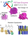
Fig. 1.
Fig. 1.
Description and models of PETase and the random copolymers. (A) GoMartini model of PETase in magenta with the active site in gray. (B) The secondary structure of PETase is shown with the same color scheme as in A. The active site is shown using the van der Waals representation to highlight the cleft-like binding pocket for PET. (C) Surface representation of PETase. (D) Chemical and Martini description of the methacrylate-based random copolymers. Hydrophobic beads are tan, while hydrophilic beads are blue, and negatively charged beads are cyan. FH, FL, and F refer to the percentage of the respective monomer in the random copolymers. (E) Snapshot of the Martini random copolymer model with the colors corresponding to D.
Fig. 2.
Fig. 2.
(A and B) Comparison of the GoMartini PETase (B) alone to atomistic simulations (A) and to the GoMartini PETase complexed with random compolymers as the temperature is increased (B). rmsd is used to measure the conformation relative to the crystal structure with higher values signifying more deformation. (A) Results for the atomistic model show a general increase in the rmsd of the protein as well as the active site as is expected based on known decreases in activity with increasing temperature. (B) The GoMartini model shows an increase in rmsd for the whole protein and the active site at 320 K, but at 350 K the active site rmsd decreases, unlike the entire protein backbone. The active site rmsd may be inaccurate at high temperatures, but can still be used as a baseline to measure the thermal stability of the PETase–polymer complexes. In these complexes, temperature dependence of the whole protein and active site conformation is nearly eliminated.
Fig. 3.
Fig. 3.
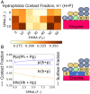
Random copolymer complexation with the PETase protein for different mean polymer compositions. FH is the percentage of hydrophobic EHMA, F is the percentage of negatively charged SPMA, and FL is the percentage of hydrophilic OEGMA-9. These percentages sum to 100 and thus FL, which is not displayed, is 100 – FHF. (A) The number of contacts is shown as a function of composition at 298 K. A maximum is observed at very low percentages of EHMA and higher percentages of SPMA, while a local minimum is observed at FH = 20%. (B) Simulation snapshot of a wrapped polymer conformation, which occurs at low values of FH and is characterized by a high percentage of contacts between the enzyme and polymer backbone. (C) Simulation snapshot of a globular polymer conformation, which occurs at high values of FH and is characterized by micelle-like behavior of the amphiphilic polymers.
Fig. 4.
Fig. 4.
Hydrophobic interactions affect PETase-random copolymer complexation. (A) The fraction of contacts that involve hydrophobic polymer beads. There are two local maxima, one at very low FH where the polymer backbone wraps around the protein surface and one at high FH where the EHMA increases the baseline hydrophobic fraction of the polymer. (B) The fraction of these hydrophobic contacts that occur on the hydrophobic surface of PETase is lower than the hydrophobic surface fraction of PETase. This struggle to optimize the interaction could be related to ill-defined hydrophobic domains due to partially hydrophobic amino acids in the Martini model. However, polar–polar interactions seem to be optimized and this increases as charge is added, while the opposite occurs for hydrophobic–hydrophobic interactions.
Fig. 5.
Fig. 5.
Including negatively charged monomers increases contact with positive surface domains. (A and B) PETase with only the surface potential and with additional contacts (ACs) overlaid, respectively. One can see that the yellow and cyan sites, which are preferred when F = 10%, are overwhelmingly on the positive part of the protein due to the addition of negative charge to the polymer. (C and D) A rotated orientation of the protein. The full dataset for this, more method description, and higher-temperature results can be found in SI Appendix, Fig. S2.
Fig. 6.
Fig. 6.
Less perturbed active sites are stabilized by additional contacts near the active site. (A) The distribution of active site rmsds at 298 and 320 K. The less perturbed active sites are colored in blue while the more perturbed active sites are colored in red. Each point refers to a different polymer composition and the line refers to PETase alone at 298 K. These are the groups being compared in C and D. (B) Surface representation of PETase with no excess contacts shown for comparison. (C) Comparison at 298 K shows many ACs around the active sites for less perturbed compositions. These contacts stabilize the active site instead of further perturbing it at 320 K (D) as well. The full dataset for this and more method description can be found in SI Appendix, Fig. S6.
Fig. 7.
Fig. 7.
Activity of PETase and PETase/copolymer complexes at two different PETase:copolymer molar ratios. (A) Specific activity against small molecule substrate p-nitrophenyl acetate after 1 h incubation at various temperatures. Error bars represent SD over three replicate experiments. (B) PET degradation activity over 5 h at 35 C. Error bars represent the 95% confidence interval on activity values.

References

    1. Coon M. J., Cytochrome P450: Nature’s most versatile biological catalyst. Annu. Rev. Pharmacol. Toxicol. 45, 1–25 (2005). - PubMed
    1. Martinez C. A., Rupashinghe S. G., Cytochrome P450 bioreactors in the pharmaceutical industry: Challenges and opportunities. Curr. Top. Med. Chem. 13, 1470–1490 (2013). - PubMed
    1. Sureka H. V., Obermeyer A. C., Flores R. J., Olsen B. D., Catalytic biosensors from complex coacervate core micelle (c3m) thin films. ACS Appl. Mater. Interfaces 11, 32354–32365 (2019). - PubMed
    1. DelRe C., et al. ., Near-complete depolymerization of polyesters with nano-dispersed enzymes. Nature 592, 558–563 (2021). - PubMed
    1. Blank L. M., Narancic T., Mampel J., Tiso T., O’Connor K., Biotechnological upcycling of plastic waste and other non-conventional feedstocks in a circular economy. Curr. Opin. Biotechnol. 62, 212–219 (2020). - PubMed

LinkOut - more resources