. 2022 Mar 21;17(3):e0265844.
doi: 10.1371/journal.pone.0265844. eCollection 2022.

Evaluation of ocular biometry in primary angle-closure disease with two swept source optical coherence tomography devices

Affiliations

Evaluation of ocular biometry in primary angle-closure disease with two swept source optical coherence tomography devices

Boonsong Wanichwecharungruang et al. PLoS One. .

Abstract

Purpose: To investigate agreement between 2 swept source OCT biometers, IOL Master700 and Anterion, in various ocular biometry and intraocular lens (IOL) calculations of primary angle-closure disease (PACD).

Setting: Rajavithi Hospital, Bangkok, Thailand.

Design: Prospective comparative study.

Methods: This study conducted in a tertiary eye care center involving biometric measurements obtained with 2 devices in phakic eye with diagnosis of PACD. Mean difference and intraclass correlation coefficient (ICC) with confidence limits were assessed, and calculations of estimated residual refraction of the IOL were analysed using Barrett's formula.

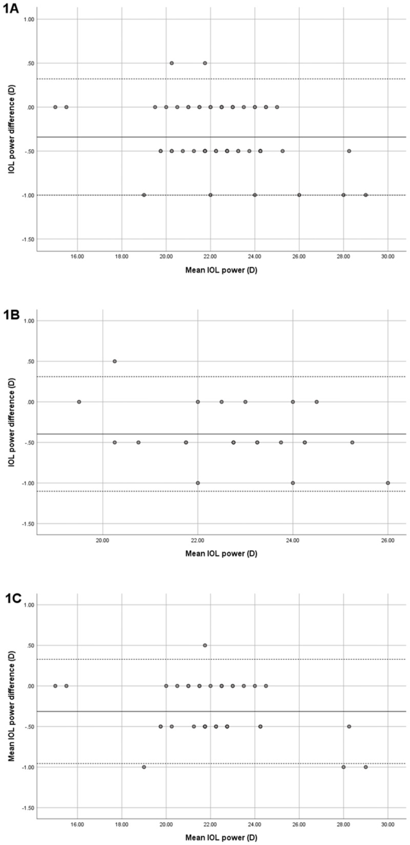
Results: Sixty-nine eyes from 45 PACD patients were enrolled for the study. Excellent agreement of various parameters was revealed, with ICC (confidence limits) of K1 = 0.953 (0.861-0.979), K2 = 0.950 (0.778-0.98), ACD = 0.932 (0.529-0.978), WTW = 0.775 (0.477-0.888), and LT = 0.947 (0.905-0.97). Mean difference of axial length (AL) was -0.01 ± 0.02 mm with ICC of 1.000. IOL calculation was assessed with Barrett's formula, and Bland-Altman plot showed excellent agreement in the results of the 2 devices for the IOL power and estimated post-operative residual refraction (EPR).

Conclusions: Mean differences of biometric parameters, obtained with IOL Master700 and Anterion, were small, and ICC showed excellent concordance. No clinical relevance in calculation of IOL power was found, and the two devices appeared to be comparably effective in clinical practice.


Conflict of interest statement

The authors have declared that no competing interests exist.

Figures

Fig 1
Fig 1. The Bland-Altman plot of the IOL power using parameters from IOLMaster700 and Anterion.
The solid line represents the mean difference, whereas dotted lines on both sides represent the upper and lower 95% LoA. A: In all PACD eyes, The plot demonstrates that only two eyes from total 69 eyes were out of the 95% LoAs, indicating excellent agreement in IOL power. B: In the eyes with ACD≤2.4 mm, The plot demonstrates that only one eye from 24 eyes were out of the 95% LoAs, indicating excellent agreement in IOL power. C: In the eyes with ACD>2.4 mm. The plot demonstrates that only one eye from 45 eyes were out of the 95% LoAs, indicating excellent agreement in IOL power.
Fig 2
Fig 2. The EPR between the devices’ measurements.
The Bland-Altman plot of the EPR using parameters from IOLMaster700 and Anterion of all PACD eyes. The solid line represents the mean difference, whereas dotted lines on both sides represent the upper and lower 95% LoA. A: In all PACD eyes, the plot demonstrates that all cases were within the 95% LoAs, indicating excellent agreement in EPR. B: In the eyes with ACD ≤ 2.4 mm, all cases were within the 95% LoAs, which indicates excellent agreement between the devices. C: In the eyes with ACD > 2.4 mm, the plot demonstrates that only one eye was out of the 95% LoAs, indicating excellent agreement between the devices.

Similar articles

Cited by

References

    1. Tham YC, Li X, Wong TY, Quigley HA, Aung T, Cheng CY. Global prevalence of glaucoma and projections of glaucoma burden through 2040: a systematic review and meta-analysis. Ophthalmology. 2014;121(11):2081–90. doi: 10.1016/j.ophtha.2014.05.013 - DOI - PubMed
    1. He M, Friedman DS, Ge J, Huang W, Jin C, Lee PS, et al.. Laser peripheral iridotomy in primary angle-closure suspects: biometric and gonioscopic outcomes: the Liwan Eye Study. Ophthalmology. 2007;114(3):494–500. doi: 10.1016/j.ophtha.2006.06.053 - DOI - PubMed
    1. Gazzard G, Foster PJ, Viswanathan AC, Devereux JG, Oen FT, Chew PTK, et al.. The severity and spatial distribution of visual field defects in primary glaucoma: a comparison of primary open-angle glaucoma and primary angle-closure glaucoma. Archives of Ophthalmology. 2002;120(12):1636–43. doi: 10.1001/archopht.120.12.1636 - DOI - PubMed
    1. Lam DS, Leung DY, Tham CC, Li FC, Kwong YY, Chiu TY, et al.. Randomized trial of early phacoemulsification versus peripheral iridotomy to prevent intraocular pressure rise after acute primary angle closure. Ophthalmology. 2008;115(7):1134–40. doi: 10.1016/j.ophtha.2007.10.033 - DOI - PubMed
    1. Wanichwecharungruang B, Phumratprapin C, Kongsomboon K, Seresirikachorn K. Real-world Surgical Outcomes of Primary Angle-closure Glaucoma. Clin Ophthalmol. 2021;15:2823–33. doi: 10.2147/OPTH.S315747 - DOI - PMC - PubMed

Publication types