. 2022 Mar 21;18(3):e1010197.
doi: 10.1371/journal.ppat.1010197. eCollection 2022 Mar.

From Deer-to-Deer: SARS-CoV-2 is efficiently transmitted and presents broad tissue tropism and replication sites in white-tailed deer

Affiliations

From Deer-to-Deer: SARS-CoV-2 is efficiently transmitted and presents broad tissue tropism and replication sites in white-tailed deer

Mathias Martins et al. PLoS Pathog. .

Abstract

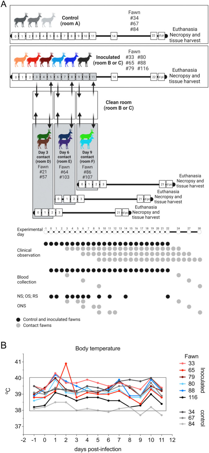
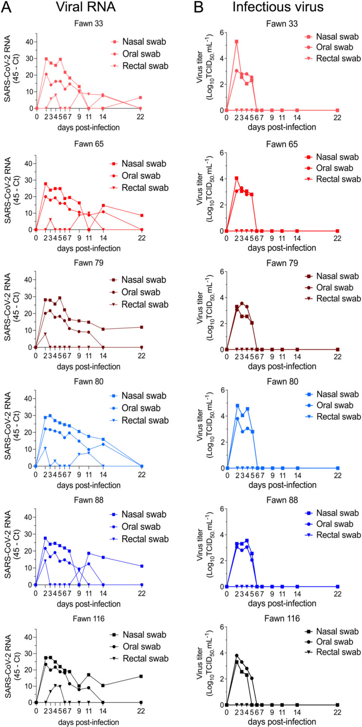
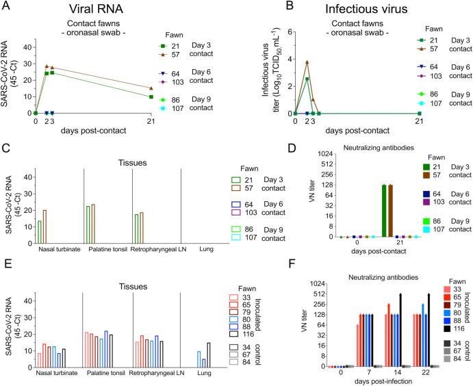
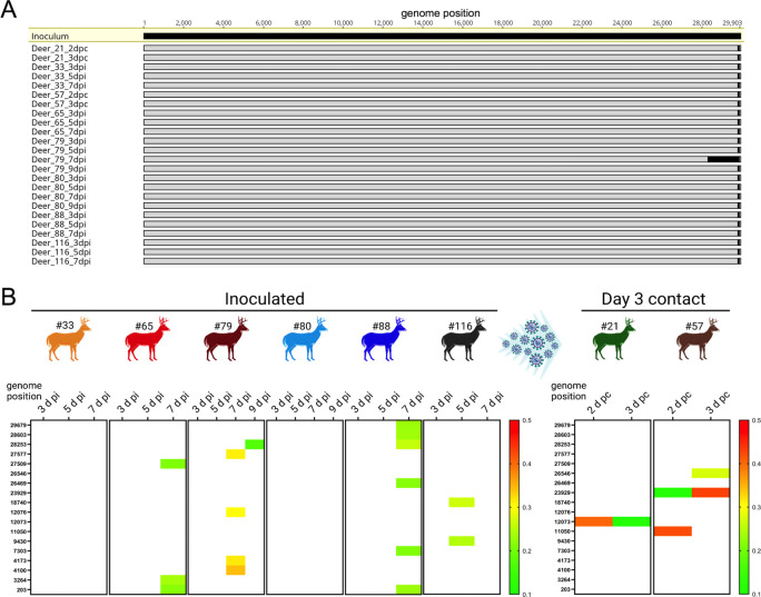
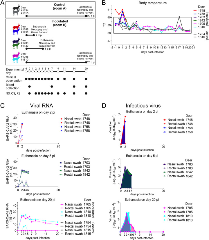
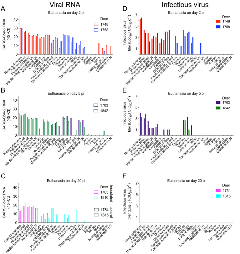
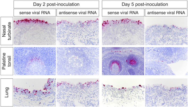
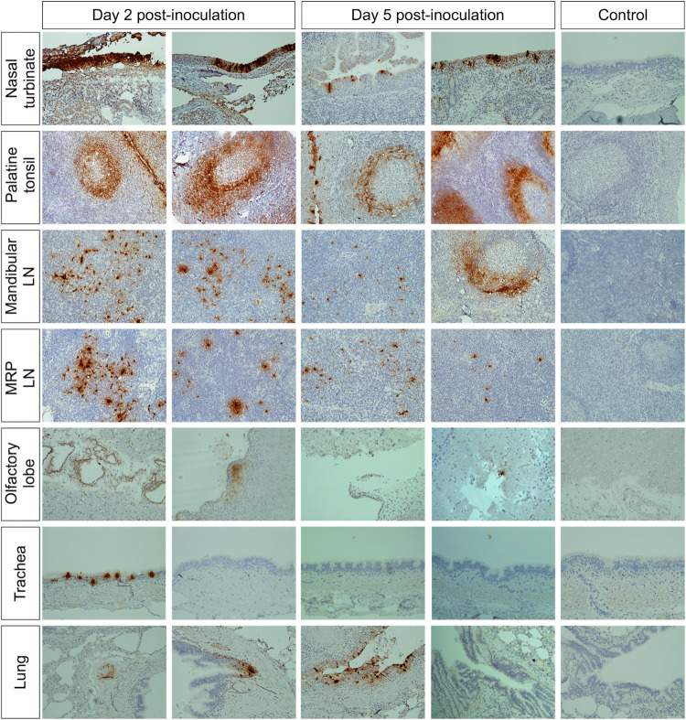
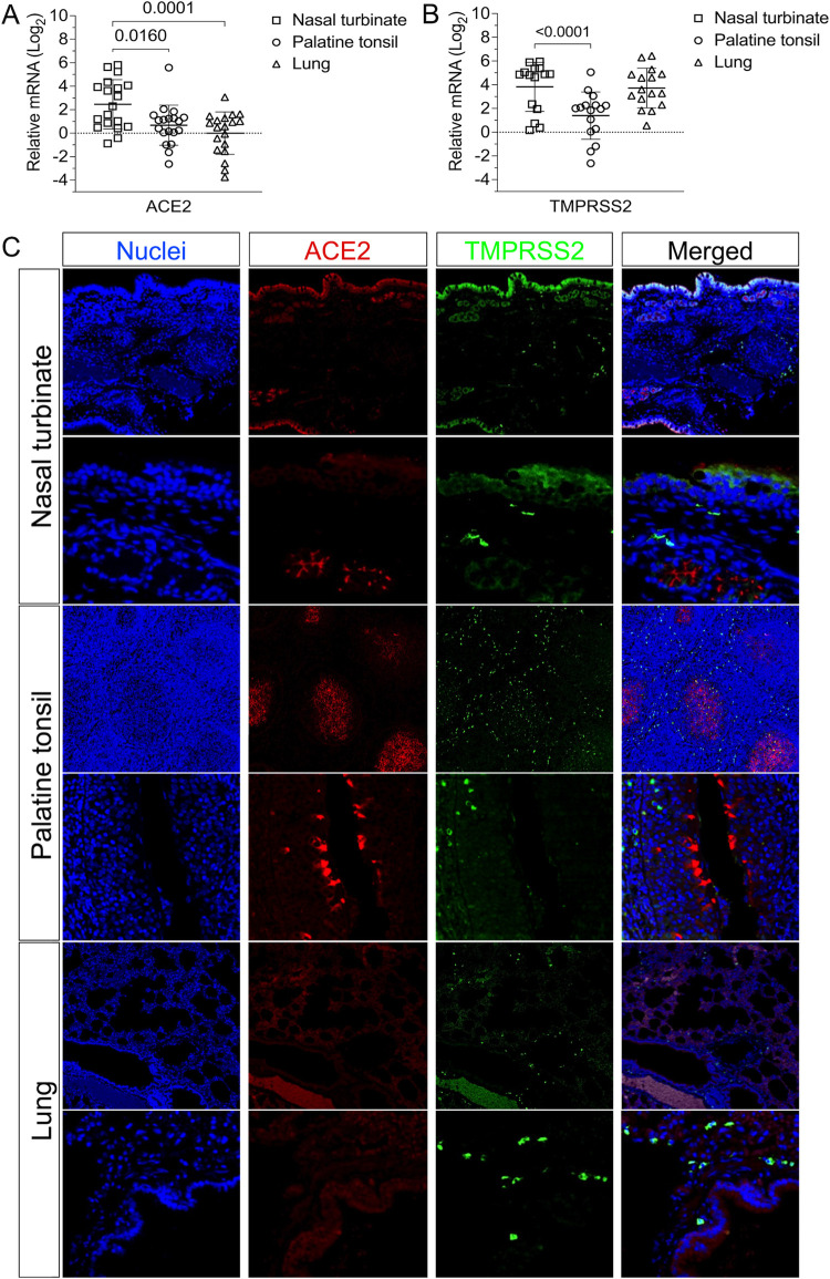
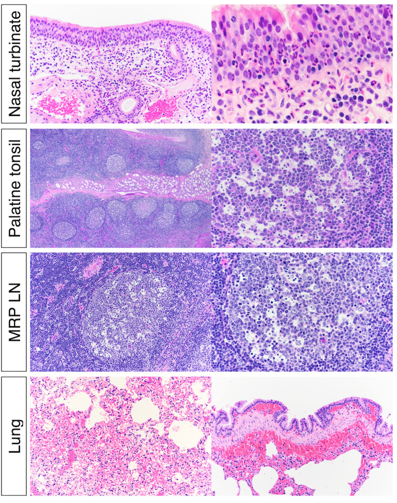
Severe acute respiratory syndrome coronavirus 2 (SARS-CoV-2), the causative agent of coronavirus disease 2019 (COVID-19) in humans, has a broad host range, and is able to infect domestic and wild animal species. Notably, white-tailed deer (WTD, Odocoileus virginianus), the most widely distributed cervid species in the Americas, were shown to be highly susceptible to SARS-CoV-2 in challenge studies and reported natural infection/exposure rates approaching 30-40% in free-ranging WTD in the U.S. Thus, understanding the infection and transmission dynamics of SARS-CoV-2 in WTD is critical to prevent future zoonotic transmission to humans, at the human-WTD interface during hunting or venison farming, and for implementation of effective disease control measures. Here, we demonstrated that following intranasal inoculation with SARS-CoV-2 B.1 lineage, WTD fawns (~8-month-old) shed infectious virus up to day 5 post-inoculation (pi), with high viral loads shed in nasal and oral secretions. This resulted in efficient deer-to-deer transmission on day 3 pi. Consistent a with lack of infectious SARS-CoV-2 shedding after day 5 pi, no transmission was observed to contact animals added on days 6 and 9 pi. We have also investigated the tropism and sites of SARS-CoV-2 replication in adult WTD (3-4 years of age). Infectious virus was detected up to day 6 pi in nasal secretions, and from various respiratory-, lymphoid-, and central nervous system tissues, indicating broad tissue tropism and multiple sites of virus replication. The study provides important insights on the infection and transmission dynamics of SARS-CoV-2 in WTD, a wild animal species that is highly susceptible to infection and with the potential to become a reservoir for the virus in the field.


Conflict of interest statement

The authors have declared that no competing interests exist.

Figures

Fig 1
Fig 1. Infection and transmission study of SARS-CoV-2 in white-tailed deer.
A) Schematic representation of the experimental design of infection/transmission study in WTD fawns. Three fawns were maintained as control (non-inoculated) in room A, and six fawns were inoculated intranasally with SARS-CoV-2 and housed in room B or C for 22 days post-inoculation (d pi). On day 3 pi two fawns (day 3 contact) were moved into a clean room (room C) where the inoculated fawns were also introduced. After 48 hours, day 3 contact fawns were transferred to a separate clean room (room D), where they were maintained until euthanized on 21 days post-contact (d pc). This process was performed with two additional fawns on day 6 pi (day 6 contact) and two fawns on day 9 pi (day 9 contact), which were housed in the separate rooms E and F, respectively. Respiratory nasal secretion, feces and serum were collected on the days indicated in the figure. B) Inoculated and control fawns were microchipped subcutaneously with temperature chips prior to the experiment, and monitored daily for body temperature starting on day 1 before inoculation until day 11 pi. Additionally, daily clinical observations were performed. Body temperatures are expressed in degrees Celsius. Rectangle highlights normal physiological body temperature range. NS = nasal swab; OS = oral swab; RS = rectal swab; ONS = oronasal swab.
Fig 2
Fig 2. Viral shedding dynamics in nasal and oral secretions and feces following SARS-CoV-2 inoculation in white-tailed deer.
A) SARS-CoV-2 RNA load assessed in nasal and oral secretions and feces of controls (animals no. 34, 67, 84, no shown) and six inoculated fawns (animals no. 33, 65, 79, 80, 88, and 116). Nasal, oral and rectal swabs collected on days 0, 2–7, 9, 11, 14, and 22 post-inoculation (pi) were tested for the presence of SARS-CoV-2 RNA by real-time reverse transcriptase PCR (rRT-PCR). B) Infectious SARS-CoV-2 load in nasal and oral secretions and feces was assessed by virus titration in RT-rPCR-positive samples. Virus titers were determined using end point dilutions and expressed as TCID50.ml-1. All the controls fawns (uninoculated) remained negative through the experimental period.
Fig 3
Fig 3. Viral load in oronasal secretions, and seroconversion in contact white-tailed deer.
A) Virus shedding (viral RNA load) assessed in oronasal secretions of day 3- (animals no. 21 and 57), day 6- (animals no. 64 and 103), and day 9 contact (animals no. 86 and 107) fawns. Oronasal swabs collected on days 0 (before contact), days 2, 3 and 21 post- contact (pc) were tested for the presence of SARS-CoV-2 RNA by real-time reverse transcriptase PCR (rRT-PCR). B) Shedding of infectious SARS-CoV-2 in oronasal secretions was assessed by virus titrations in rRT-PCR-positive samples. Virus titers were determined using end point dilutions and expressed as TCID50.ml-1. C) Tissue distribution of SARS-CoV-2 RNA assessed in nasal turbinate, palatine tonsil, retropharyngeal lymph node, and lung collected and processed for rRT-PCR 21 d pc of contact fawns. D) Antibody responses following contact of fawns with inoculated animals was assessed by virus neutralization (VN) assay. Neutralizing antibody titers were expressed as the reciprocal of the highest dilution of serum that completely inhibited SARS-CoV-2 infection/replication. E) Tissue distribution of SARS-CoV-2 RNA assessed in nasal turbinate, palatine tonsil, retropharyngeal lymph node, and lung collected and processed for rRT-PCR 22 days post-inoculation (d pi). F) Antibody responses following SARS-CoV-2 inoculation assessed by VN assay. Neutralizing antibody titers were expressed as the reciprocal of the highest dilution of serum that completely inhibited SARS-CoV-2 infection/replication.
Fig 4
Fig 4. Low genetic diversity observed following SARS-CoV-2 replication and transmission in white-tailed deer.
Genome sequences were obtained directly from nasal secretions from all the 6 inoculated fawns collected on days 3, 5, 7 and/or 9 post-inoculation (pi) and from oronasal secretions from the day 3 contact animals (n = 2) collected on days 2 and 3 post-contact (pc). A) Whole genome sequence analyses of SARS-CoV-2 sequences recovered from all inoculated and contact fawns revealed no amino acid differences in the consensus SARS-CoV-2 genome in comparison to the genome sequence of the inoculum virus isolate NYI67-20. B) Minor variant viral populations distributed throughout the virus genome from inoculated fawns on days 3, 5, 7 and/or 9 pi and from oronasal secretions from the day 3 contact animals (n = 2) collected on days 2 and 3 pc.
Fig 5
Fig 5. Pathogenesis and tissue tropism of SARS-CoV-2 in white-tailed deer.
A) Schematic representation of the pathogenesis study design. Adult deer were kept in two rooms of a biosafety level 3 (agriculture) (BSL-3Ag) facility. Two deer were maintained as control (uninoculated) in a separate rom (room A), and six deer were inoculated intranasally with 5 x 106.38 TCID50 of SARS-CoV-2 isolate NYI67-20 (lineage B1) and housed in room B. To assess viral tissue tropism, two deer were euthanized on day 2 post-inoculation (pi) (animals no.1748 and 1758), two on day 5 pi (animals no. 1703 and 1842), and two inoculated (animals no. 1705 and 1810) and two control (animals no. 1754 and 1815) deer were euthanized on day 20 pi. B) Inoculated and control deer were microchipped subcutaneously for temperature monitoring. Temperature and clinical signs were monitored daily starting on day 1 before inoculation until day 20 pi or until euthanasia. Body temperatures are expressed in degrees Celsius. C-E) SARS-CoV-2 RNA load assessed in respiratory secretions and feces by real-time reverse transcriptase PCR (rRT-PCR) in deer until days 2 (C), 5 (D), or 20 pi (E). F-H) Infectious SARS-CoV-2 assessed in respiratory secretions and feces assessed by virus titration in rRT-PCR-positive samples until days 2 (F), 5 (G), or 20 pi (H). Virus titers were determined using end point dilutions and expressed as TCID50.ml-1. All control deer (uninoculated) remained negative throughout the experimental period.
Fig 6
Fig 6. Tissue distribution of SARS-CoV-2 RNA and infectious virus in white-tailed deer.
Tissues were collected and processed for real-time reverse transcriptase PCR (rRT-PCR) and virus titrations. A-C) Tissue distribution of SARS-CoV-2 RNA assessed in twenty-four tissues collected and processed for rRT-PCR in deer on days 2 (A), 5 (B), or 20 post-inoculation (pi) (C). D-F) Infectious SARS-CoV-2 in tissues assessed by virus titrations in RT-rPCR-positive samples obtained on days 2 (D), 5 (E) or 20 pi (F). Virus titers were determined using end point dilutions and expressed as TCID50.ml-1. All the controls fawns (uninoculated) remained negative through the experimental period.
Fig 7
Fig 7. In situ hybridization (ISH) in tissues from white-tailed deer inoculated with SARS-CoV-2.
Paraffin-embedded tissues were subjected to ISH using the RNAscope ZZ probe technology. Nasal turbinate, palatine tonsil and lung from deer on days 2 and 5 post-inoculation (pi). Intense labeling of viral RNA (all viral RNA) highlighted on the three tissues on days 2 and 5 pi. Labeling using the antisense genome probe demonstrate genome RNA replication in the nasal turbinate, palatine tonsil, and lung on day 2 pi and less intense on day 5 pi.
Fig 8
Fig 8. Immunohistochemistry (IHC) in tissues from white-tailed deer inoculated with SARS-CoV-2.
SARS-CoV-2 labeling in the tissues of the inoculated WTD by IHC showing staining for the SARS-CoV-2 N protein (brown stain) in several tissues on days 2 and 5 post-inoculation (pi). Tissue sections were counter stained with hematoxylin. Tissues from a control (uninoculated) animal were included in all IHC stains.
Fig 9
Fig 9. Expression of ACE2 and TMPRSS2 in the tissues of white-tailed deer.
A-B) The levels of ACE2 and TMPRSS2 gene transcription in nasal turbinate, palatine tonsil and lungs were assessed by quantitative real-time reverse transcriptase PCR (qRT-PCR). Tissues from all 15 fawns from the transmission study (euthanized on days 21 or 22) and 4 deer from the pathogenesis study (euthanized on day 20) were included. Expression levels of ACE2 (A) and TMPRSS2 (B) in nasal turbinate, palatine tonsil and lung. C) Expression of ACE2 and TMPRSS2 proteins in the nasal turbinate, palatine tonsil, and lung are presented. Paraffin-embedded tissues from a control deer (uninoculated) were subjected to an immunofluorescence assay using a polyclonal antibody anti-ACE2 (red) and a monoclonal antibody anti-TMPRSS2 (green). Nuclear counterstain was performed with DAPI (blue).
Fig 10
Fig 10. Histological examination of nasal turbinate from white-tailed deer inoculated with SARS-CoV-2.
Upper respiratory tract (URT) after inoculation, there was submucosal lymphoplasmacytic infiltrate and frequent mucosal exudation of neutrophils in nasal turbinate. Numerous germinal centers of the palatine tonsils were characterized by lymphoid depletion and lymphocytolysis. Within the lung there was diffuse congestion with multifocal submucosal hemorrhages associated with larger bronchi.

References

    1. Gorbalenya AE, Baker SC, Baric RS, de Groot RJ, Drosten C, Gulyaeva AA, et al.. The species Severe acute respiratory syndrome-related coronavirus: classifying 2019-nCoV and naming it SARS-CoV-2. Nature Microbiology. Nature Research; 2020. pp. 536–544. doi: 10.1038/s41564-020-0695-z - DOI - PMC - PubMed
    1. Zhou P, Yang X Lou, Wang XG, Hu B, Zhang L, Zhang W, et al.. A pneumonia outbreak associated with a new coronavirus of probable bat origin. Nature. 2020;579: 270–273. doi: 10.1038/s41586-020-2012-7 - DOI - PMC - PubMed
    1. Murakami S, Kitamura T, Suzuki J, Sato R, Aoi T, Fujii M, et al.. Detection and Characterization of Bat Sarbecovirus Phylogenetically Related to SARS-CoV-2, Japan. Emerg Infect Dis. 2020. doi: 10.3201/eid2612.203386 - DOI - PMC - PubMed
    1. Lau SKP, Luk HKH, Wong ACP, Li KSM, Zhu L, He Z, et al.. Possible Bat Origin of Severe Acute Respiratory Syndrome Coronavirus 2. Emerg Infect Dis. 2020;26: 1542–1547. doi: 10.3201/eid2607.200092 - DOI - PMC - PubMed
    1. Temmam S, Vongphayloth K, Salazar EB, Munier S, Bonomi M, Régnault B, et al.. Coronaviruses with a SARS-CoV-2-like receptor- binding domain allowing ACE2-mediated entry into human cells isolated from bats of Indochinese peninsula. Res Sq. 2021.

Supplementary concepts