

Fully Integrated Ultra-thin Intraoperative Micro-imager for Cancer Detection Using Upconverting Nanoparticles
- PMID: 35312938
- PMCID: PMC9970948
- DOI: 10.1007/s11307-022-01710-8
Fully Integrated Ultra-thin Intraoperative Micro-imager for Cancer Detection Using Upconverting Nanoparticles
Abstract
Purpose: Intraoperative detection and removal of microscopic residual disease (MRD) remain critical to the outcome of cancer surgeries. Today's minimally invasive surgical procedures require miniaturization and surgical integration of highly sensitive imagers to seamlessly integrate into the modern clinical workflow. However, current intraoperative imagers remain cumbersome and still heavily dependent on large lenses and rigid filters, precluding further miniaturization and integration into surgical tools.
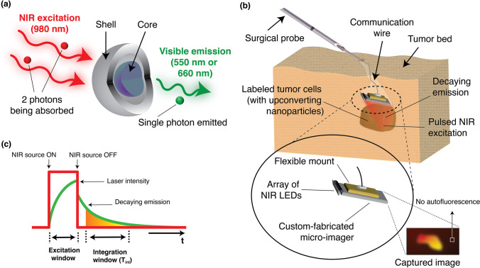
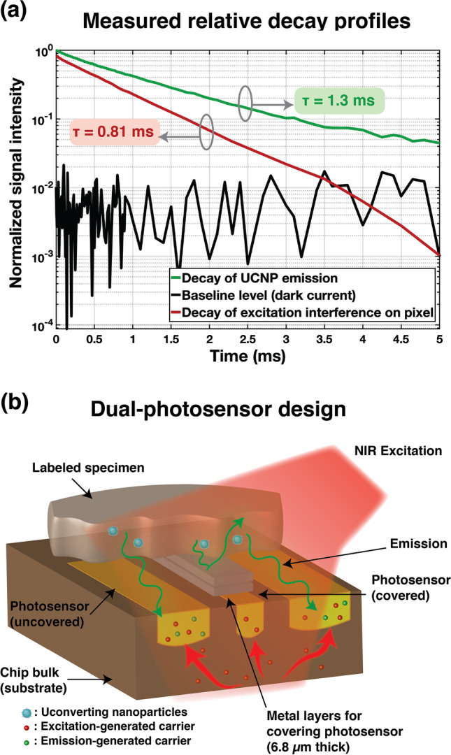
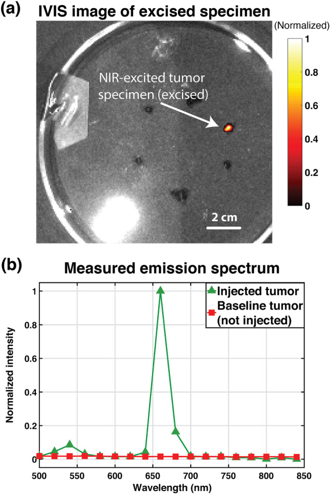
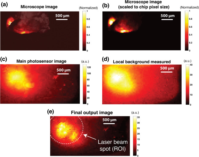
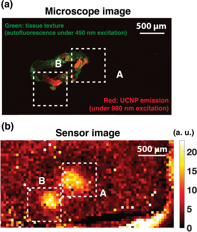
Procedures: We have successfully engineered a chip-scale intraoperative micro-imager array-without optical filters or lenses-integrated with lanthanide-based alloyed upconverting nanoparticles (aUCNPs) to achieve tissue imaging using a single micro-chip. This imaging platform is able to leverage the unique optical properties of aUCNPs (long luminescent lifetime, high-efficiency upconversion, no photobleaching) by utilizing a time-resolved imaging method to acquire images using a 36-by-80-pixel, 2.3 mm [Formula: see text] 4.8 mm silicon-based electronic imager micro-chip, that is, less than 100-µm thin. Each pixel incorporates a novel architecture enabling automated background measurement and cancellation. We have validated the performance, spatial resolution, and the background cancellation scheme of the imaging platform, using resolution test targets and mouse prostate tumor sample intratumorally injected with aUCNPs. To demonstrate the ability to image MRD, or tumor margins, we evaluated the imaging platform in visualizing a single-cell thin section of the injected prostate tumor sample.
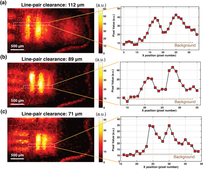
Results: Tested on USAF resolution targets, the imager is able to achieve a resolution of 71 µm. We have also demonstrated successful background cancellation, achieving a signal-to-background ratio of 8 when performing ex vivo imaging on aUCNP-injected prostate tumor sample, improved from originally 0.4. The performance of the imaging platform on single-cell layer sections was also evaluated and the sensor achieved a signal-to-background ratio of 4.3 in resolving cell clusters with sizes as low as 200 cells.
Conclusion: The imaging system proposed here is a scalable chip-scale ultra-thin alternative for bulky conventional intraoperative imagers. Its novel pixel architecture and background correction scheme enable visualization of microscopic-scale residual disease while remaining completely free of lenses and filters, achieving an ultra-miniaturized form factor-critical for intraoperative settings.
Keywords: Intraoperative microscopy; Silicon imager; Time-resolved imaging; Upconverting nanoparticle.
© 2022. The Author(s).
Conflict of interest statement
The authors declare no competing interests.
Figures
Similar articles
-
A 25 micron-thin microscope for imaging upconverting nanoparticles with NIR-I and NIR-II illumination.Theranostics. 2019 Oct 18;9(26):8239-8252. doi: 10.7150/thno.37672. eCollection 2019. Theranostics. 2019. PMID: 31754393 Free PMC article.
-
Optics-Free Chip-Scale Intraoperative Imaging Using NIR-Excited Upconverting Nanoparticles.IEEE Trans Biomed Circuits Syst. 2022 Apr;16(2):312-323. doi: 10.1109/TBCAS.2022.3165186. Epub 2022 May 19. IEEE Trans Biomed Circuits Syst. 2022. PMID: 35385388 Free PMC article.
-
A Molecular Imaging "Skin A Time-resolving Intraoperative Imager for Microscopic Residual Cancer Detection Using Enhanced Upconverting Nanoparticles.Annu Int Conf IEEE Eng Med Biol Soc. 2018 Jul;2018:1-4. doi: 10.1109/EMBC.2018.8512947. Annu Int Conf IEEE Eng Med Biol Soc. 2018. PMID: 30440277
-
Optimizing brain tumor resection. Midfield interventional MR imaging.Neuroimaging Clin N Am. 2001 Nov;11(4):659-72. Neuroimaging Clin N Am. 2001. PMID: 11995421 Review.
-
Future perspectives for intraoperative MRI.Neurosurg Clin N Am. 2005 Jan;16(1):201-13. doi: 10.1016/j.nec.2004.07.011. Neurosurg Clin N Am. 2005. PMID: 15561539 Review.
Cited by
-
Multicolor fluorescence microscopy for surgical guidance using a chip-scale imager with a low-NA fiber optic plate and a multi-bandpass interference filter.Biomed Opt Express. 2024 Feb 20;15(3):1761-1776. doi: 10.1364/BOE.509235. eCollection 2024 Mar 1. Biomed Opt Express. 2024. PMID: 38495694 Free PMC article.
-
Editorial to the Special Issue Entitled "Optical Surgical Navigation".Mol Imaging Biol. 2023 Feb;25(1):1-2. doi: 10.1007/s11307-023-01806-9. Mol Imaging Biol. 2023. PMID: 36729349 No abstract available.
-
Optical Coherence Tomography Angiography of the Intestine: How to Prevent Motion Artifacts in Open and Laparoscopic Surgery?Life (Basel). 2023 Mar 6;13(3):705. doi: 10.3390/life13030705. Life (Basel). 2023. PMID: 36983861 Free PMC article.
References
-
- Buchholz TA, Somerfield MR, Griggs JJ, et al. Margins for breast-conserving surgery with whole-breast irradiation in stage I and II invasive breast cancer: American society of clinical oncology endorsement of the society of surgical oncology/American society for radiation oncology consensus guideline. J Clin Oncol. 2014;32:1502–1506. doi: 10.1200/JCO.2014.55.1572. - DOI - PubMed
-
- Moran MS, Schnitt SJ, Giuliano AE, et al. Society of Surgical Oncology-American Society for Radiation Oncology Consensus Guideline on Margins for Breast-Conserving Surgery With Whole-Breast Irradiation in Stages I and II Invasive Breast Cancer. In J Radiat Oncol Biol Phys. 2014;88:553–564. doi: 10.1016/J.IJROBP.2013.11.012. - DOI - PMC - PubMed
