Review
. 2022 Mar;16(1):1-18.
doi: 10.1007/s12105-021-01406-5. Epub 2022 Mar 21.

Update from the 5th Edition of the World Health Organization Classification of Head and Neck Tumors: Nasal Cavity, Paranasal Sinuses and Skull Base

Affiliations
Review

Update from the 5th Edition of the World Health Organization Classification of Head and Neck Tumors: Nasal Cavity, Paranasal Sinuses and Skull Base

Lester D R Thompson et al. Head Neck Pathol. 2022 Mar.

Abstract

The World Health Organization Classification of Head and Neck Tumours recently published the 5th edition. There are new entities, emerging entities, and significant updates to the taxonomy and characterization of tumor and tumor-like lesions, specifically in this article as it relates to nasal cavity, paranasal sinuses and skull base. Importantly, the number of diagnostic entries has been reduced by creating category-specific chapters for soft tissue, hematolymphoid, melanocytic, neuroectodermal, and metastatic tumors. Bone and salivary gland tumors are also not separately reported in the sinonasal tract, but included in the jaw and salivary gland sections, respectively. Repetition of characteristic entities in each anatomic site was also reduced, instead highlighting only the unique features in each anatomic site. Two new entities (SWI/SNF complex-deficient sinonasal carcinomas and HPV-related multiphenotypic sinonasal carcinoma) will be highlighted in this review, with a discussion of several emerging entities. There is a short description of updated information for all 24 diagnostic entities included in this edition to allow the reader a snapshot of current state of knowledge, but to encourage more investigation and further broaden understanding of these diverse and rare entities.

Keywords: Carcinoma; Immunohistochemistry; Nasal cavity; Papillomavirus neoplasms; Paranasal sinus neoplasms; Paranasal sinuses; SWI/SNF complex; Skull base; World Health Organization.


Conflict of interest statement

All authors have contributed to this work and both authors declare they have no conflict of interest as it relates to this study.

Figures

Fig. 1
Fig. 1
Sinonasal hamartomas. A Respiratory epithelial adenomatoid hamartoma. An axial computed tomography shows expanded olfactory cleft (white arrow) with a soft tissue-density mass. B Evenly spaced glandular units have a prominent basement membrane surrounding them. C. Seromucinous hamartoma shows a proliferation of small eosinophilic glands in the stroma, lacking destructive growth. D A chondromesenchymal hamartoma shows cartilaginous nodules with a myxoid stroma and islands of bony tissue (courtesy Dr. D. Baumhoer)
Fig. 2
Fig. 2
Nonkeratinizing SCC. A A computed tomography scan demonstrates a soft tissue mass involving the nasal cavity and maxillary sinus, with bone erosion. B Tumor invasion is as smooth-edged lobules and ribbons, reminiscent of inverted papilloma. C This case harbored DEK::AFF2. Cases with this fusion typically have nuclear monotony and in infiltrate of neutrophils. D Nonkeratinizing SCC is characteristically positive for p40 in a diffuse pattern
Fig. 3
Fig. 3
SMARCB1-deficient sinonasal carcinoma. A Most cases have a basaloid low-power appearance. Bone invasion is common. B A minority of cases have a “pink” cell appearance, where the tumor cells are somewhat plasmacytoid, with hyaline-appearing cytoplasm. C Rare cases show glandular or even yolk sac-like differentiation. D A complete loss of SMARCB1 expression by immunohistochemistry defines this tumor. Note the retained staining in lymphocytes, a helpful internal control
Fig. 4
Fig. 4
Sinonasal undifferentiated carcinoma (SNUC). A SNUC is an aggressive tumor, commonly showing invasion of local structures like the orbit or brain. B A nonspecific, basaloid, nested appearance is typical. C The tumor cells are monotonous with necrosis and a high mitotic rate. D SNUC is usually completely negative for squamous markers like p40
Fig. 5
Fig. 5
Teratocarcinosarcoma. A) There is a blend of spindled cells, epithelial elements and primitive cells. B) The mesenchymal spindled cell component is juxtaposed with malignant glandular elements. C) An area of squamoid differentiation shows clear cell change, while the primitive neuroectodermal component has a high nuclear to cytoplasmic ratio. D SMARCA4 is lost as reflected by a negative BRG1 stain. Note retained expression in lymphocytes, a helpful internal control
Fig. 6
Fig. 6
HPV-related multiphenotypic sinonasal carcinoma. A The tumor infiltrates as basaloid nests and strands within a myxoid stroma. Surface epithelial dysplasia is seen overlying the tumor. B Myoepithelial cell markers like SMA are positive in an abluminal pattern. C CD117 highlights tumor ducts which are often subtle on routine microscopy. D The presence of high-risk HPV required for a diagnosis of HPV-related multiphenotypic sinonasal carcinoma. In this case, it was demonstrated by RNA in situ hybridization
Fig. 7
Fig. 7
Low-grade non-intestinal-type sinonasal adenocarcinoma. A At low-power, there is a markedly increased number of seromucinous glands within the nasal submucosa. B The tumor glands have minimal cellular atypia, but demonstrate architectural atypia in the form of fusion and cribriforming with no intervening stroma. C A rare variant known as renal cell carcinoma-like adenocarcinoma has water-clear cytoplasm and prominent cell membranes. D S100 protein is usually positive, supporting seromucinous differentiation
Fig. 8
Fig. 8
Biphenotypic sinonasal sarcoma. A The right-sided tumor involves the nasal cavity with bone erosion. B The tumor is infiltrative and often entraps downward invaginations of surface respiratory-type epithelium. C The tumor grows as fascicles of uniform spindle cells with minimal mitotic activity or atypia. Slit-like vessels are common. D In some cases the tumor can show overt rhabdomyoblastic differentiation in the form of strap cells
Fig. 9
Fig. 9
Sinonasal ameloblastoma. A An axial CT shows an ethmoid sinus mass expanding and filling the sinus with a soft tissue-density mass. B A central stellate reticulum with ameloblastic palisade. C The surface respiratory epithelium overlies the proliferation of ameloblastoma. D An intact, ciliated respiratory-type epithelium is seen overlying a proliferation of columnar, basaloid cells associated with a well-developed central stellate reticulum
Fig. 10
Fig. 10
Adamantinomatous craniopharyngioma. A A destructive skull base mass extends into the nasopharynx (white arrow). B A solid and cystic proliferation of stellate reticulum and anucleated squamous epithelium. C A cellular stellate reticulum with numerous calcifications (black arrow). D Wet keratin is identified as anucleated squamous cell adjacent to the epithelium

References

    1. Bishop JA, Loney EL, Thompson LDR. Nasal cavity, paranasal sinuses, and skull base tumours. In: Board WCTE, editor. Head and neck tumours. 5. Lyon: International Agency for Research on Cancer; 2022.
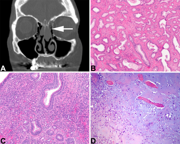
    1. Ozolek JA, Hunt JL. Tumor suppressor gene alterations in respiratory epithelial adenomatoid hamartoma (REAH): comparison to sinonasal adenocarcinoma and inflamed sinonasal mucosa. Am J Surg Pathol. 2006;30:1576–1580. - PubMed
    1. Baneckova M, Michal M, Laco J, et al. Immunohistochemical and genetic analysis of respiratory epithelial adenomatoid hamartomas and seromucinous hamartomas: are they precursor lesions to sinonasal low-grade tubulopapillary adenocarcinomas? Hum Pathol. 2020;97:94–102. - PubMed
    1. Lee JT, Garg R, Brunworth J, Keschner DB, Thompson LD. Sinonasal respiratory epithelial adenomatoid hamartomas: series of 51 cases and literature review. Am J Rhinol Allergy. 2013;27:322–328. - PubMed
    1. Daniel A, Wong E, Ho J, Singh N. Chondro-osseous respiratory epithelial adenomatoid hamartoma (COREAH): case report and literature review. Case Rep Otolaryngol. 2019;2019:5247091. - PMC - PubMed

LinkOut - more resources