Review
. 2022 Mar;16(1):54-62.
doi: 10.1007/s12105-021-01402-9. Epub 2022 Mar 21.

Update from the 5th Edition of the World Health Organization Classification of Head and Neck Tumors: Tumours of the Oral Cavity and Mobile Tongue

Affiliations
Review

Update from the 5th Edition of the World Health Organization Classification of Head and Neck Tumors: Tumours of the Oral Cavity and Mobile Tongue

Susan Muller et al. Head Neck Pathol. 2022 Mar.

Abstract

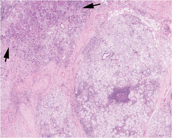
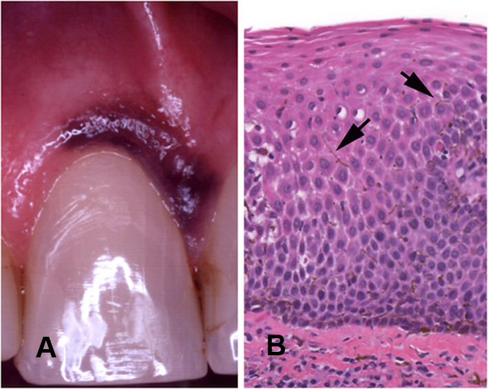
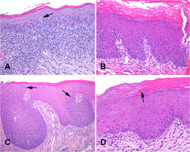
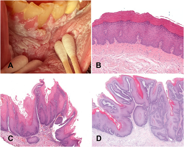
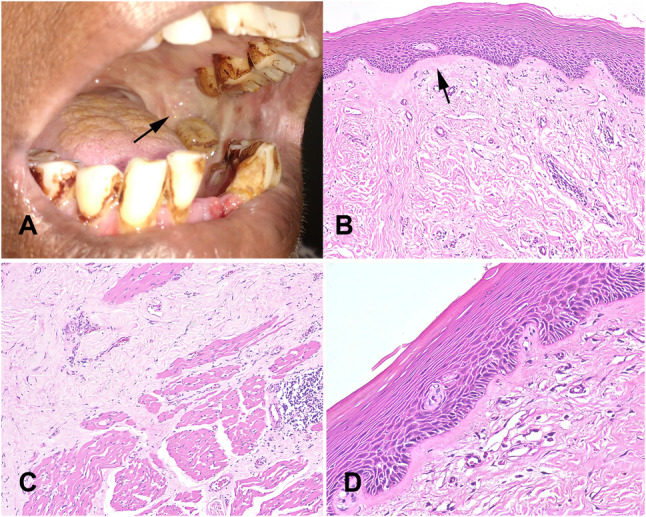
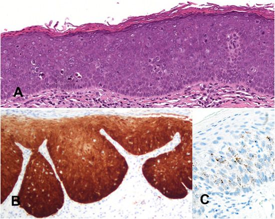
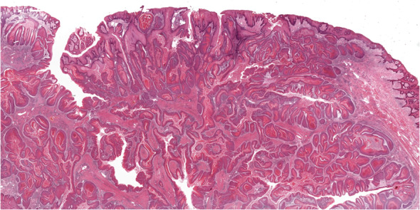
The fifth chapter of the upcoming fifth edition of the 2022 World Health Organization Classification of Tumours of the Head and Neck titled Tumours of the oral cavity and mobile tongue, has had some modifications from the 2017 fourth edition. A new section "Non-neoplastic Lesions", introduces two new entries: necrotizing sialometaplasia and melanoacanthoma. The combined Oral potentially malignant disorders and Oral epithelial dysplasia section in the 2015 WHO has now been separated and submucous fibrosis and HPV-associated dysplasia are also discussed in separate sections. Carcinoma cuniculatum and verrucous carcinoma are described in dedicated sections, reflecting that the oral cavity is the most common location in the head and neck for both these entities which have distinct clinical and histologic features from conventional squamous cell carcinoma. This review summarizes the changes in Chapter 5 with special reference to new additions, deletions, and sections that reflect current clinical, histological, and molecular advances.

Keywords: Dysplasia; Ectomesenchymal chondromyxoid tumor; Mobile tongue; Oral cavity; Proliferative verrucous leukoplakia; Squamous cell; Submucous fibrosis; World Health Organization.


Conflict of interest statement

Neither author has any conflicts of interest to disclose.

Figures

Fig. 1
Fig. 1
Necrotizing sialometaplasia showing the outline of residual acini with necrosis with preservation of the lobular architecture of the glands and squamous metaplasia of residual ducts (arrows). Courtesy or Dr. K Magliocca
Fig. 2
Fig. 2
Melanoacanthoma. A Diffuse melanin pigment affecting the attached gingiva of the maxillary central incisor. B Acanthotic epithelium with dendritic melanocytes (arrows) within the intercellular spaces
Fig. 3
Fig. 3
Oral epithelial dysplasia. A Moderate dysplasia with a band-like inflammatory infiltrate at the epithelial-stromal interface imparting a lichenoid appearance but with general preservation of a slightly expanded and hyperchromatic basal cell layer. A dyskeratotic cell (arrow) is present in the spinous layer. B Mild dysplasia showing primarily an architectural or differentiated pattern of dysplasia with marked orthohyperkeratosis, loss of basal cell polarization, budding of rete, increased hyperchromasia, and mitotic figures confined to basal and parabasal layer. C Skip areas of dysplasia (arrows) indicate a clonal pattern which can be present in HPV-associated dysplasia. This case was negative for HPV16 by in situ hybridization. D Sharp demarcation from normal parakeratotic epithelium to orthokeratotic epithelium in mild epithelial dysplasia. The small basal cells give rise to cells with eosinophilic cytoplasm without cytologic atypia. This pattern of dysplasia is primarily an architectural pattern
Fig. 4
Fig. 4
Proliferative verrucous leukoplakia. A An advanced case with a large area of leukoplakia involving the labial mucosa, mandibular vestibule and attached gingiva. B Early lesion exhibiting subtle histology with orthokeratosis, prominent granular layer, and no cytologic atypia. C A late stage of PVL showing acanthosis and a well-developed papillary architecture with a lichenoid immune response. D Bulky hyperkeratotic exophytic and endophytic growth pattern covered by parakeratin with an undulating surface and surface crypts filled with parakeratin
Fig. 5
Fig. 5
Submucous fibrosis A. Leathery mucosa, blanching and trismus (arrow). Teeth show betel quid staining. B Atrophic epithelium and dense fibrosis of the corium with juxta-epithelial hyalinization (arrow). C Progressive replacement of muscle by fibrous tissue. D Epithelial dysplasia starts to appear with budding rete morphology in the atrophic epithelium
Fig. 6
Fig. 6
HPV-associated dysplasia. A Epithelium with a brightly eosinophilic keratin surface and basaloid morphology showing marked architectural and cytological changes of dysplasia with karyorrhectic and apoptotic cells. B Full thickness diffuse and intense cytoplasmic and nuclear immunoreactivity for p16. C RNA in situ hybridization for high-risk HPV shows punctate dot-like nuclear and cytoplasmic staining
Fig. 7
Fig. 7
Carcinoma cuniculatum showing the characteristic infiltrative labyrinthine network of keratin-filled crypts. Courtesy of Dr. Amrita Jay

References

    1. WHO Classification of Tumours Editorial Board. Head and neck tumours (WHO classification of tumours series, 5th ed. vol. 9). Lyon (France): International Agency for Research on Cancer 2022.
    1. Carlson DL. Necrotizing sialometaplasia: a practical approach to the diagnosis. Arch Pathol Lab Med. 2009;133(5):692–698. - PubMed
    1. Abrams AM, Melrose RJ, Howell FV. Necrotizing sialometaplasia. A disease simulating malignancy. Cancer. 1973;32(1):130–135. - PubMed
    1. Fowler CB, Brannon RB. Subacute necrotizing sialadenitis: report of 7 cases and a review of the literature. Oral Surg Oral Med Oral Pathol Oral Radiol Endod. 2000;89(5):600–609. - PubMed
    1. Kaplan I, et al. The clinical, histologic, and treatment spectrum in necrotizing sialometaplasia. Oral Surg Oral Med Oral Pathol Oral Radiol. 2012;114(5):577–585. - PubMed

MeSH terms