Review
. 2022 Mar;16(1):123-142.
doi: 10.1007/s12105-022-01435-8. Epub 2022 Mar 21.

Update from the 5th Edition of the World Health Organization Classification of Head and Neck Tumors: Overview of the 2022 WHO Classification of Head and Neck Neuroendocrine Neoplasms

Affiliations
Review

Update from the 5th Edition of the World Health Organization Classification of Head and Neck Tumors: Overview of the 2022 WHO Classification of Head and Neck Neuroendocrine Neoplasms

Ozgur Mete et al. Head Neck Pathol. 2022 Mar.

Abstract

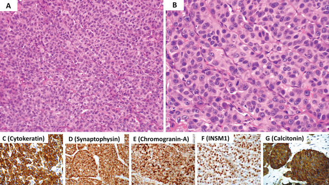
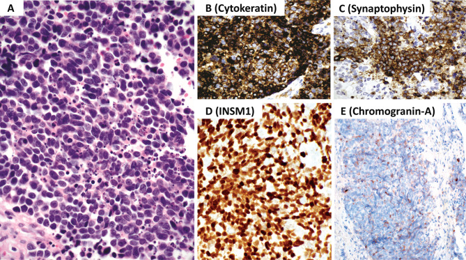
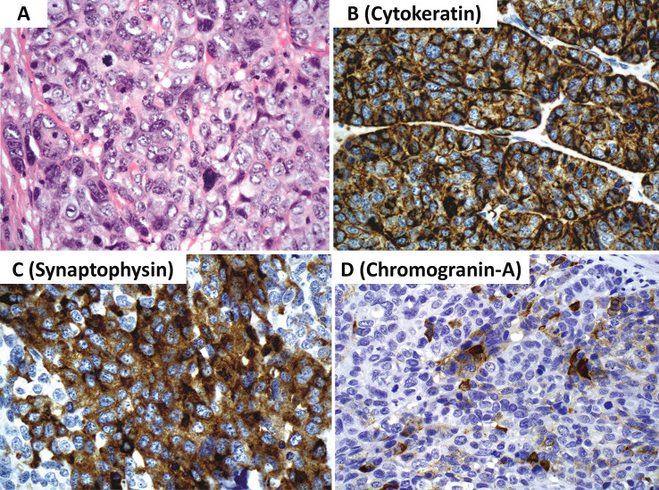
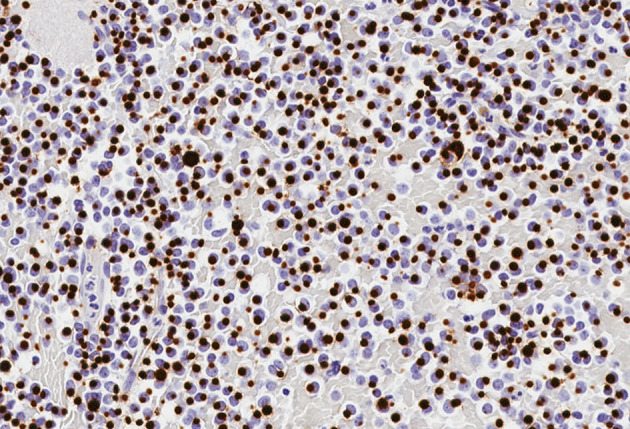
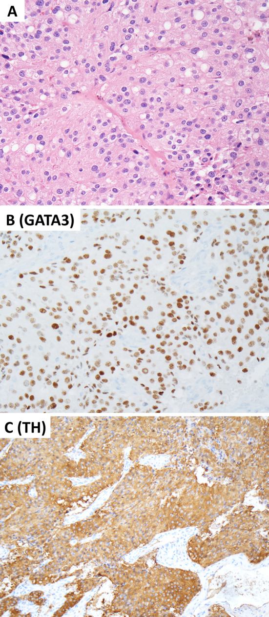
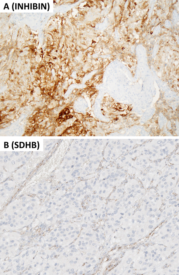
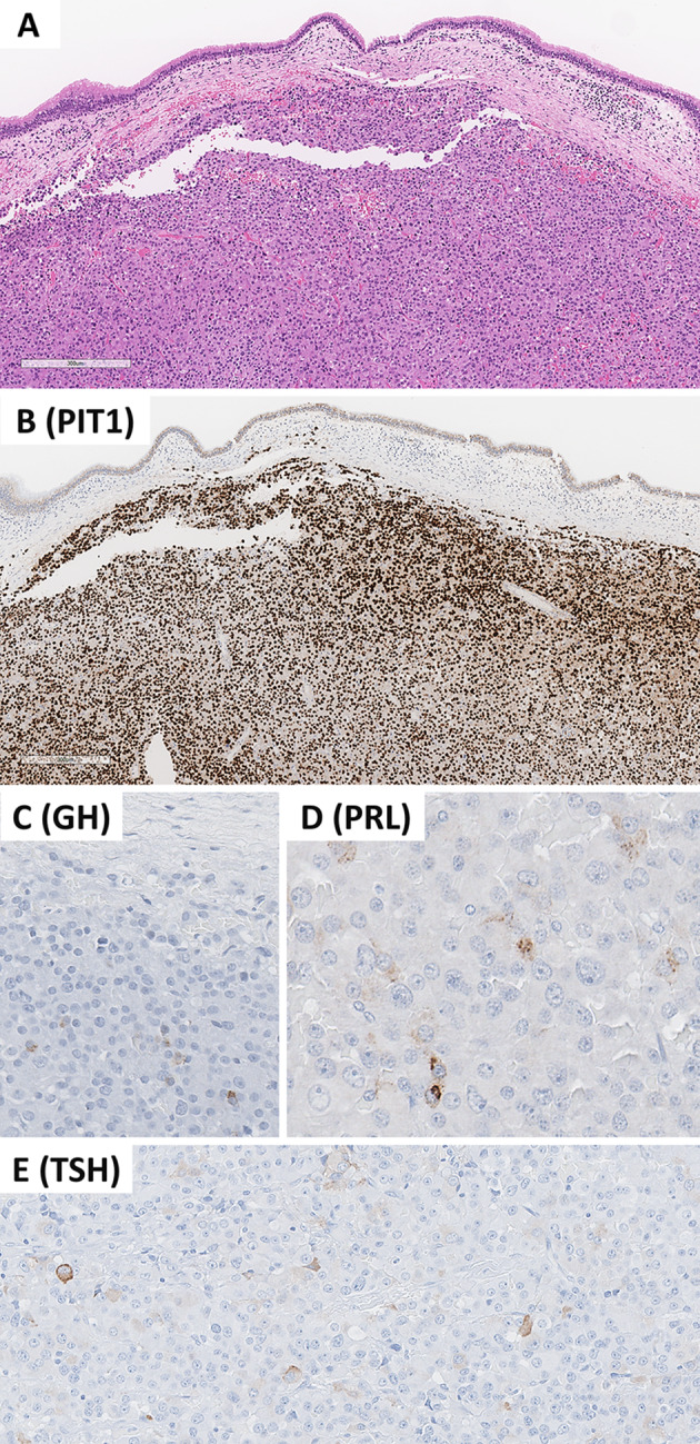
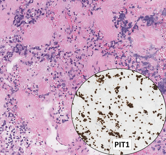
This review article provides a brief overview of the new WHO classification by adopting a question-answer model to highlight the spectrum of head and neck neuroendocrine neoplasms which includes epithelial neuroendocrine neoplasms (neuroendocrine tumors and neuroendocrine carcinomas) arising from upper aerodigestive tract and salivary glands, and special neuroendocrine neoplasms including middle ear neuroendocrine tumors (MeNET), ectopic or invasive pituitary neuroendocrine tumors (PitNET; formerly known as pituitary adenoma) and Merkel cell carcinoma as well as non-epithelial neuroendocrine neoplasms (paragangliomas). The new WHO classification follows the IARC/WHO nomenclature framework and restricts the diagnostic term of neuroendocrine carcinoma to poorly differentiated epithelial neuroendocrine neoplasms. In this classification, well-differentiated epithelial neuroendocrine neoplasms are termed as neuroendocrine tumors (NET), and are graded as G1 NET (no necrosis and < 2 mitoses per 2 mm2; Ki67 < 20%), G2 NET (necrosis or 2-10 mitoses per 2 mm2, and Ki67 < 20%) and G3 NET (> 10 mitoses per 2 mm2 or Ki67 > 20%, and absence of poorly differentiated cytomorphology). Neuroendocrine carcinomas (> 10 mitoses per 2 mm2, Ki67 > 20%, and often associated with a Ki67 > 55%) are further subtyped based on cytomorphological characteristics as small cell and large cell neuroendocrine carcinomas. Unlike neuroendocrine carcinomas, head and neck NETs typically show no aberrant p53 expression or loss of RB reactivity. Ectopic or invasive PitNETs are subtyped using pituitary transcription factors (PIT1, TPIT, SF1, GATA3, ER-alpha), hormones and keratins (e.g., CAM5.2). The new classification emphasizes a strict correlation of morphology and immunohistochemical findings in the accurate diagnosis of neuroendocrine neoplasms. A particular emphasis on the role of biomarkers in the confirmation of the neuroendocrine nature of a neoplasm and in the distinction of various neuroendocrine neoplasms is provided by reviewing ancillary tools that are available to pathologists in the diagnostic workup of head and neck neuroendocrine neoplasms. Furthermore, the role of molecular immunohistochemistry in the diagnostic workup of head and neck paragangliomas is discussed. The unmet needs in the field of head and neck neuroendocrine neoplasms are also discussed in this article. The new WHO classification is an important step forward to ensure accurate diagnosis that will also form the basis of ongoing research in this field.

Keywords: Biomarkers; Head and neck neuroendocrine neoplasms; Merkel cell carcinoma; Neuroendocrine carcinoma; Neuroendocrine tumors; Paraganglioma; Pituitary adenoma; Pituitary neuroendocrine tumor; WHO classification.


Conflict of interest statement

The authors declare that this manuscript was conducted in the absence of any commercial or financial relationships that could be construed as a potential conflict of interest.

Figures

Fig. 1
Fig. 1
Middle ear neuroendocrine tumor (MeNET) with low grade features (G1 MeNET). A Low magnification shows a combination n of growth patterns including trabecular, ribbons, glandular and solid. B The neoplasm is composed of cuboidal to columnar cells with round to oval nuclei, dispersed (“salt and pepper”) nuclear chromatin, inconspicuous nucleoli and eosinophilic cytoplasm. C Cells with plasmacytoid features are commonly present. The is an absence of significant nuclear pleomorphism, increased mitotic activity and/or tumor necrosis. The neoplastic cells are diffusely immunoreactive for D cytokeratin using the AE1/AE3 and CAM5.2 cocktail, E synaptophysin, F chromogranin-A and G INSM1. These tumors often show L-cell differentiation (immunoreactive for peptide-YY, glucagon and pancreatic polypeptide) along with SATB2 expression (not illustrated herein). Contributor: Dr. Bruce Wenig
Fig. 2
Fig. 2
Laryngeal neuroendocrine tumor with intermediate grade features (G2 NET). A and B The neoplasm has organoid and trabecular growth composed of epithelioid cells with round to oval nuclei, dispersed nuclear chromatin, inconspicuous to identifiable nucleoli and eosinophilic cytoplasm. The is a mild to moderate nuclear pleomorphism, and increased mitotic activity, the latter ranges from 2 to 10 mitoses per 2 mm2. Tumor necrosis may also be present although not shown here. The neoplastic cells are diffusely immunoreactive for C cytokeratin using the AE1/AE3 and CAM5.2 cocktail), D synaptophysin, E chromogranin-A, F INSM1 and G calcitonin. Contributor: Dr. Bruce Wenig
Fig. 3
Fig. 3
Sinonasal neuroendocrine carcinoma, small cell type. A The neoplasm has sheetlike growth and is composed of small cells (smaller than the diameter of 3 lymphocytes), hyperchromatic to stippled appearing nuclear chromatin, inconspicuous nucleoli and scant cytoplasm. Nuclear molding, apoptosis and increased mitotic activity is present. The mitotic count should reach a level of greater than 10 mitoses per 2 mm2. B The neoplastic cells are immunoreactive for cytokeratin (AE1/AE3 and CAM5.2 cocktail). C The tumor cells are near diffusely positive for chromogranin-A and/or synaptophysin. D Diffuse INSM1 is required to confirm the diagnosis when diffuse chromogranin-A is not identified. E Most tumors may have limited to absent chromogranin-A reactivity. Contributor: Dr. Bruce Wenig
Fig. 4
Fig. 4
Sinonasal neuroendocrine carcinoma, large cell type. A Large cell neuroendocrine carcinoma is composed of cells larger than the diameter of 3 lymphocytes with round to oval nuclei, vesicular chromatin, prominent nucleoli and eosinophilic cytoplasm. Characteristic growth patterns of neuroendocrine neoplasms (not shown) as well as marked nuclear pleomorphism, increased mitotic activity (should reach a level of greater than 10 mitoses per 2 mm2) and tumor necrosis (not shown) are also a part of the diagnostic criteria along with a Ki67 > 20% (often way over 50%). B The neoplastic cells are immunoreactive for cytokeratin (AE1/AE3 and CAM5.2 cocktail). C There is often near diffuse synaptophysin expression. D Chromogranin-A staining but be very limited or even absent. Diffuse INSM1 (not illustrated herein) is required to confirm the diagnosis when diffuse chromogranin-A is not identified. Contributor: Dr. Bruce Wenig
Fig. 5
Fig. 5
Fibrous bodies in a sparsely granulated somatotroph tumor. This photomicrograph illustrates juxta-nuclear globular CAM5.2 reactivity (fibrous bodies) in an invasive sparsely granulated somatotroph tumor. This tumor is also positive for PIT1 (not illustrated) and is focally weakly positive for GH (not illustrated). Contributor: Dr. Ozgur Mete
Fig. 6
Fig. 6
Crooke cell tumor. Crooke cell tumor is an aggressive subtype of TPIT-lineage PiTNETs. A The tumor cells show Crooke’s hyaline change. PAS and ACTH highlight secretory granules which are typically dislocated to the cell periphery and juxtanuclear area. B The tumor cells show ring-like diffuse perinuclear reactivity, characteristics of these tumors. C Consistent with their corticotroph origin, these tumors are diffusely positive for TPIT. Contributor: Dr. Ozgur Mete
Fig. 7
Fig. 7
Pathologist-driven automated image analysis nuclear algorithm in the assessment of the Ki67 labeling index. Contributor: Dr. Ozgur Mete
Fig. 8
Fig. 8
Head and Neck Paraganglioma. This composite figure illustrates a carotid body paraganglioma with a mixed dopaminergic and noradrenergic secretory phenotype. The tumor arises in a patient with a germline pathogenic SDHB variant. A The tumor cells show variable eosinophilic cytoplasm with vacuoles consistent with the morphological features of SDHx-related pathogenesis. These non-epithelial neuroendocrine neoplasms are positive for GATA3 (B) and tyrosine hydroxylase (C). Contributor: Dr. Ozgur Mete
Fig. 9
Fig. 9
Molecular immunohistochemistry in head and neck paraganglioma. This composite figure illustrates a SDH-deficient head and neck paraganglioma. A Alpha-inhibin expression occurs frequently in pseudohypoxia pathway-related paragangliomas including those with SDHx-alterations. B The same tumor shows global loss of cytoplasmic granular SDHB reactivity while the endothelial cells (nontumorous elements) remain positive. This finding warrants a diagnosis of SDH-deficient paraganglioma, and requires germline SDHx (SDHA, SDHB, SDHC, SDHD, and SDHAF2) testing. Contributor: Dr. Ozgur Mete
Fig. 10
Fig. 10
Invasive pituitary neuroendocrine tumor (PitNET). This composite photomicrograph illustrates an invasive PitNET consisting of an immature PIT1 lineage tumor that manifested with a mass occupying lesion in the sphenoid sinus. The involvement of the pituitary gland on imaging studies excluded the possibility of ectopic PitNET. The tumor involves the respiratory mucosa (A) and show positive reactivity for PIT1 (B). Unlike mature forms of PIT1-lineage tumors, these tumors show focal/variable reactivity for one or more than one PIT1-lineage hormones including growth hormone (C), prolactin (D), and beta-TSH (E). Contributor: Dr. Ozgur Mete
Fig. 11
Fig. 11
Lactotroph tumor with dopamine agonist therapy. This photomicrograph illustrates an invasive sparsely granulated lactotroph tumor with extensive fibrosis. The tumor cells have scant cytoplasm and they may be mistaken for lymphoid cells; however, diffuse PIT1 reactivity confirms the diagnosis of PitNET. The tumor cells are also positive for ER-alpha and prolactin (focal/dot-like). Contributor: Dr. Ozgur Mete
Fig. 12
Fig. 12
Merkel cell carcinoma. Merkel cell carcinomas are poorly differentiated cutaneous neuroendocrine carcinomas. These tumors are positive for chromogranin-A (B) and CK20 with variable dot-like reactivity (C). They are associated with increased MIB1/KI67 proliferation index (D). Positivity for MCPyV using the CM2B4 antibody is seen in most tumors (E). Positivity for p63 is associated with adverse biology (F). Contributor: Dr. Ozgur Mete

References

    1. Rosai J. The origin of neuroendocrine tumors and the neural crest saga. Mod Pathol. 2011;24:S53–S57. - PubMed
    1. Pearse AG. The cytochemistry and ultrastructure of polypeptide hormone-producing cells of the APUD series and the embryologic, physiologic and pathologic implications of the concept. J Histochem Cytochem. 1969;17:303–313. - PubMed
    1. Fontaine J, Le Douarin NM. Analysis of endoderm formation in the avian blastoderm by use of quail-chick chimeras. The problem of the neuroectodermal origin of the cells of the APUD series. J Embryol Exp Morphol. 1977;41:209–222. - PubMed
    1. Oberndorfer S. Karzinoide Tumoren des Dünndarms. Frankf Z Pathol. 1907;1:426–432.
    1. Gosset A, Masson P. Tumeurs endocrines de lappendice. Presse Med. 1914;25:237–240.