. 2022 May 18;23(10):e202200076.
doi: 10.1002/cbic.202200076. Epub 2022 Mar 30.

Conformationally Constrained Sialyl Analogues as New Potential Binders of h-CD22

Affiliations

Conformationally Constrained Sialyl Analogues as New Potential Binders of h-CD22

Rosa Ester Forgione et al. Chembiochem. .

Erratum in

Abstract

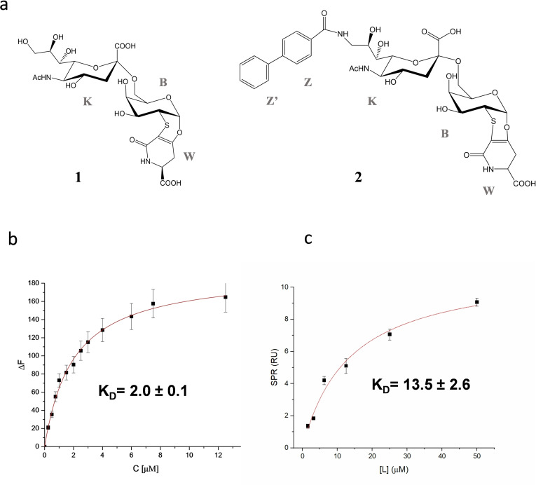
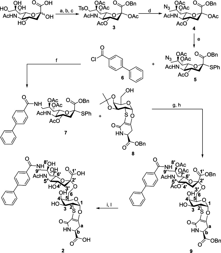
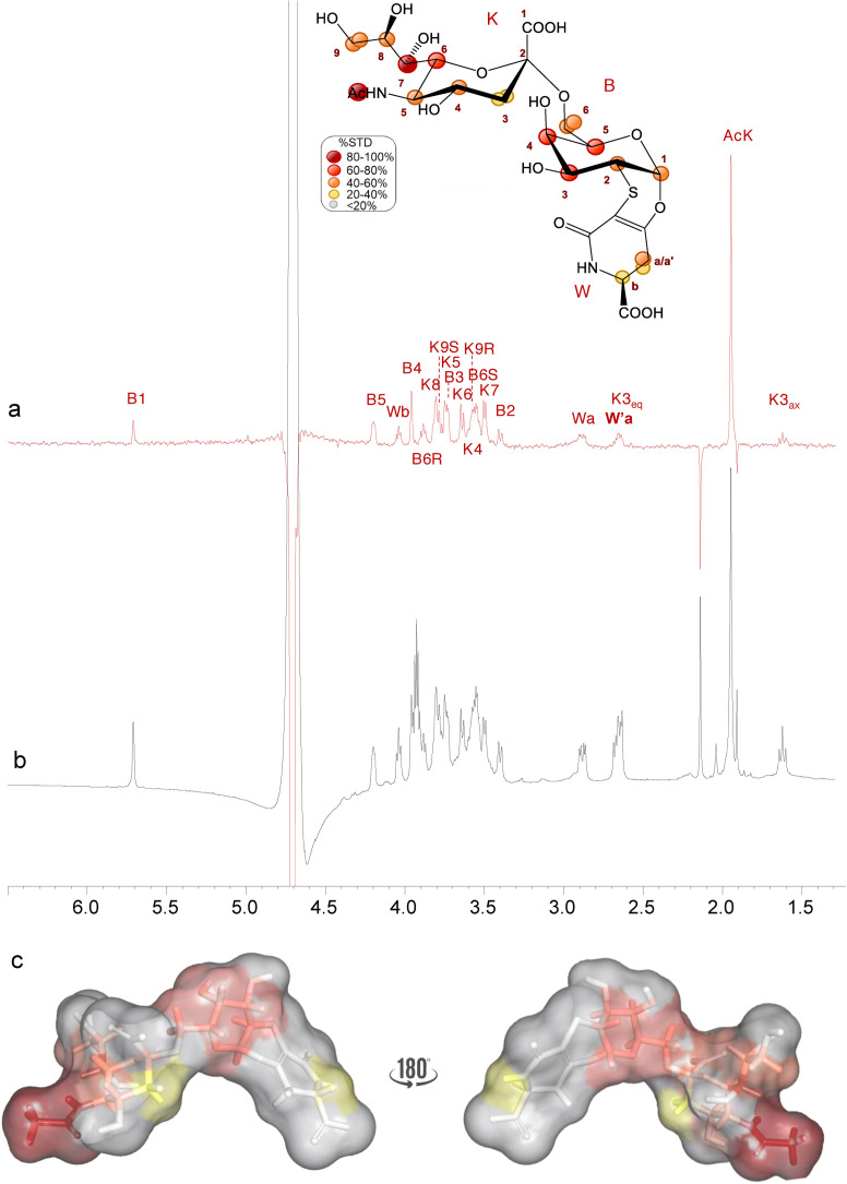
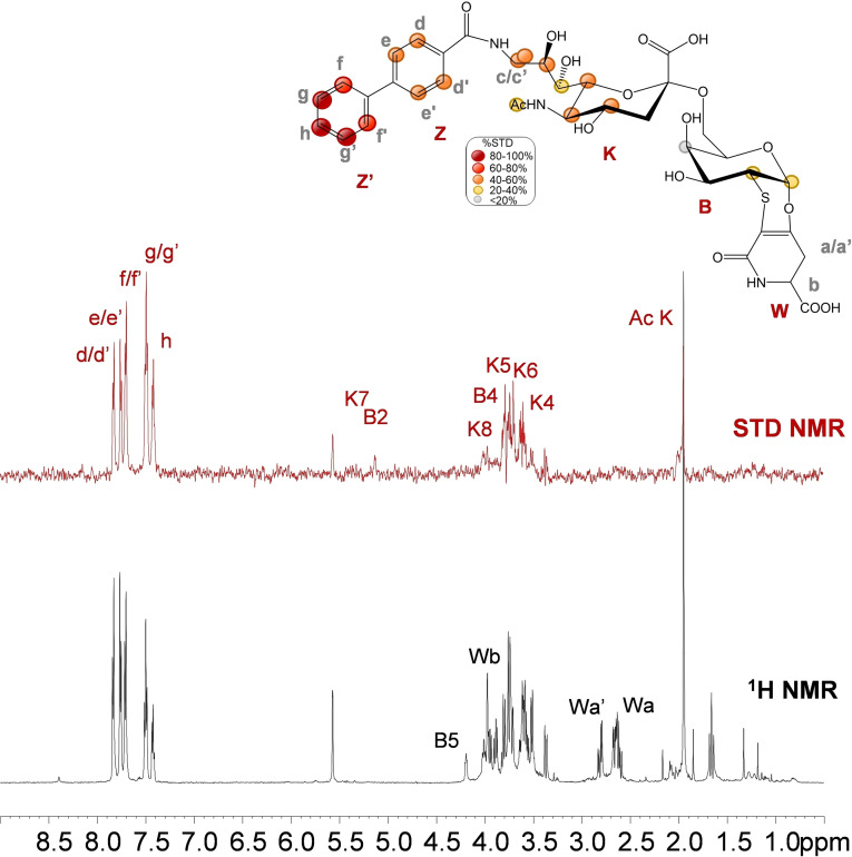
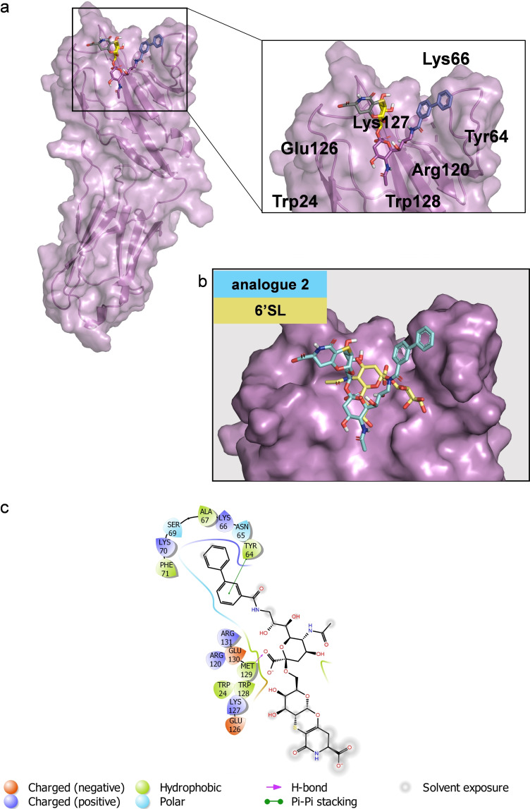
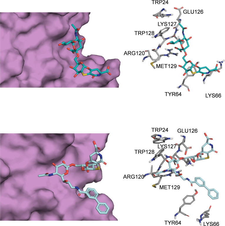
Here, two conformationally constrained sialyl analogues were synthesized and characterized in their interaction with the inhibitory Siglec, human CD22 (h-CD22). An orthogonal approach, including biophysical assays (SPR and fluorescence), ligand-based NMR techniques, and molecular modelling, was employed to disentangle the interaction mechanisms at a molecular level. The results showed that the Sialyl-TnThr antigen analogue represents a promising scaffold for the design of novel h-CD22 inhibitors. Our findings also suggest that the introduction of a biphenyl moiety at position 9 of the sialic acid hampers canonical accommodation of the ligand in the protein binding pocket, even though the affinity with respect to the natural ligand is increased. Our results address the search for novel modifications of the Neu5Ac-α(2-6)-Gal epitope, outline new insights for the design and synthesis of high-affinity h-CD22 ligands, and offer novel prospects for therapeutic intervention to prevent autoimmune diseases and B-cell malignancies.

Keywords: NMR spectroscopy; Siglecs; glycans; h-CD22; molecular recognition.


Conflict of interest statement

The authors declare no conflict of interest.

Figures

Figure 1
Figure 1
Binding affinity of h‐CD22 and sialic acid analogues. a) Structures of the sialic acid analogues used in this study. b) Fluorescence titration of h‐CD22 upon the addition of analogue 1. Each emission spectrum was recorded at the excitation wavelength of 285 nm and a temperature of 10 °C. The relative binding isotherm and the value of the dissociation constant (KD) is reported. For each data point, 10 % Y error bars are shown. c) SPR binding curve for analogue 2 vs h‐CD22 on Protein A. Each point is representative of 3 replicates (RUmean±SD).
Scheme 1
Scheme 1
Synthesis of sialo derivative 2 with atom labelling. Reagents and conditions: (a) BnBr, DBU, DMF, room temperature, 18 h; (b) TsCl, Py, room temperature, 18 h; (c) Ac2O, DMAP, Py, room temperature, 5 h; (d) NaN3, dry DMF, 70 °C, 5 h; (e) PhSH, BF3⋅Et2O, dry DCM, room temperature, 18 h; (f) PPh3, CH2Cl2, room temperature, 48 h; (g) NIS, TfOH, dry CH3CN/CH2Cl2 (10 : 1), −40 °C, 4 h; (h) AcOH 80 %, 40 °C, 18 h; (i) Pd/C, H2, MeOH, room temperature, 72 h; (l) NH3 4 M in MeOH, room temperature, 120 h.
Figure 2
Figure 2
STD NMR analysis of analogue 1 in the interaction with h‐CD22. Superimposition of the STD NMR spectrum (a) and the 1H NMR spectrum (b) of h‐CD22/analogue 1 mixture with a molecular ratio of 1 : 100, at 298 K. The interacting epitope map of analogue 1 as derived by STD‐NMR data is also reported. c) 3D representation of the analogue 1 in the bioactive conformation obtained by tr‐NOESY with molecular surface colored according to STD enhancements.
Figure 3
Figure 3
Interaction between h‐CD22 and analogue 1 by molecular modelling. a) 3D model derived by docking and MD simulations for the analogue 1 bound to h‐CD22 (PDB ID: 5VKM). The representative frame of the most populated MD cluster, obtained by Kmeans algorithm, was considered to depict the complex. b) Superimposition of the previously obtained X‐ray complex of h‐CD22/6’sialyllactose (6’SL) and the analogue 1 bound model. c) Two‐dimensional plots representing the interactions between the analogue 1 and the binding site residues of h‐CD22.
Figure 4
Figure 4
STD NMR analysis of analogue 2 in the interaction with CD22. Interacting epitope map of analogue 2 as derived by STD‐NMR data (top panel). 1H NMR and STD NMR spectra of h‐CD22/analogue 2 mixture with a molecular ratio of 1 : 100, at 298 K (bottom panel).
Figure 5
Figure 5
Interaction between CD22 and analogue 2 by molecular modelling. a) 3D model derived by docking calculations of 2 bound to h‐CD22 (PDB ID: 5VKM). The lowest energy cluster binding mode was considered to depict the complex. b) Superimposition of the previously obtained X‐ray complex of h‐CD22/6’SL and the analogue 2 bound model. c) Two‐dimensional plots representing the interactions between the analogue 2 and the binding site residues of h‐CD22.
Figure 6
Figure 6
Comparison of the binding modes of analogues 1 and 2 upon interaction with h‐CD22. Close up view of the analogues 1 (light blue sticks) and 2 (cyan sticks) into the h‐CD22 binding site (violet molecular surface). The h‐CD22 residues binding to the ligands are represented as grey sticks.

References

    1. Schnaar R. L., J. Leukocyte Biol. 2016, 99, 825–838. - PMC - PubMed
    1. Müller J., Nitschke L., Nat. Rev. Rheumatol. 2014, 10, 422–428. - PubMed
    1. Duan S., Paulson J. C., Annu. Rev. Immunol. 2020, 38, 365–395. - PubMed
    1. Crocker P. R., Varki A., Immunology 2011, 103, 137–145. - PMC - PubMed
    1. Pillai S., Netravali I. A., Cariappa A., Mattoo H., Annu. Rev. Immunol. 2012, 30, 357–392. - PMC - PubMed

Publication types

Substances