. 2022 May:51:102291.
doi: 10.1016/j.redox.2022.102291. Epub 2022 Mar 17.

The synthetic triterpenoids CDDO-TFEA and CDDO-Me, but not CDDO, promote nuclear exclusion of BACH1 impairing its activity

Affiliations

The synthetic triterpenoids CDDO-TFEA and CDDO-Me, but not CDDO, promote nuclear exclusion of BACH1 impairing its activity

Laura Casares et al. Redox Biol. 2022 May.

Abstract

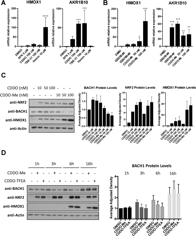
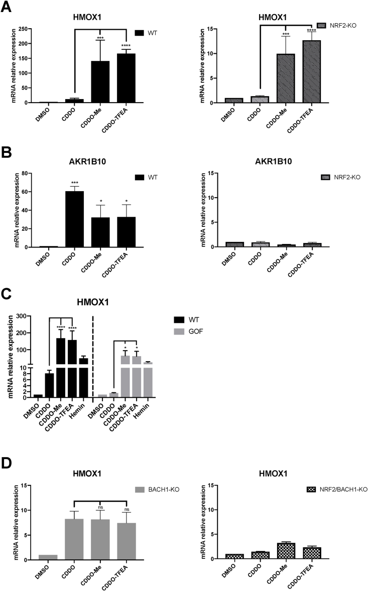
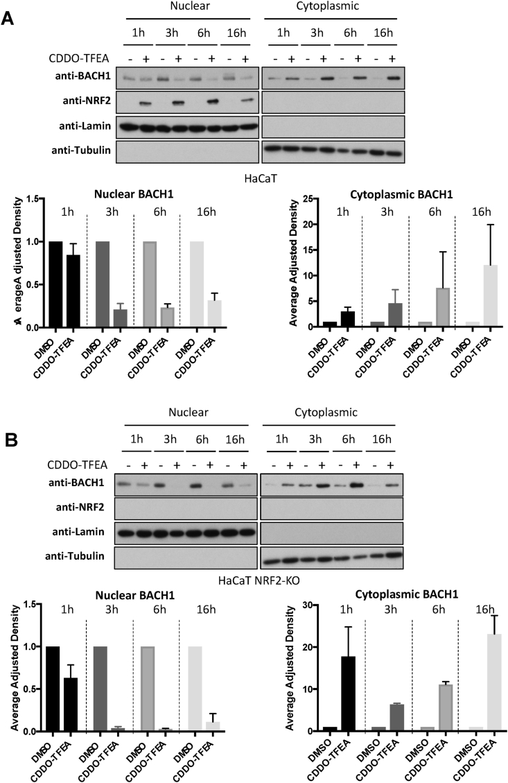
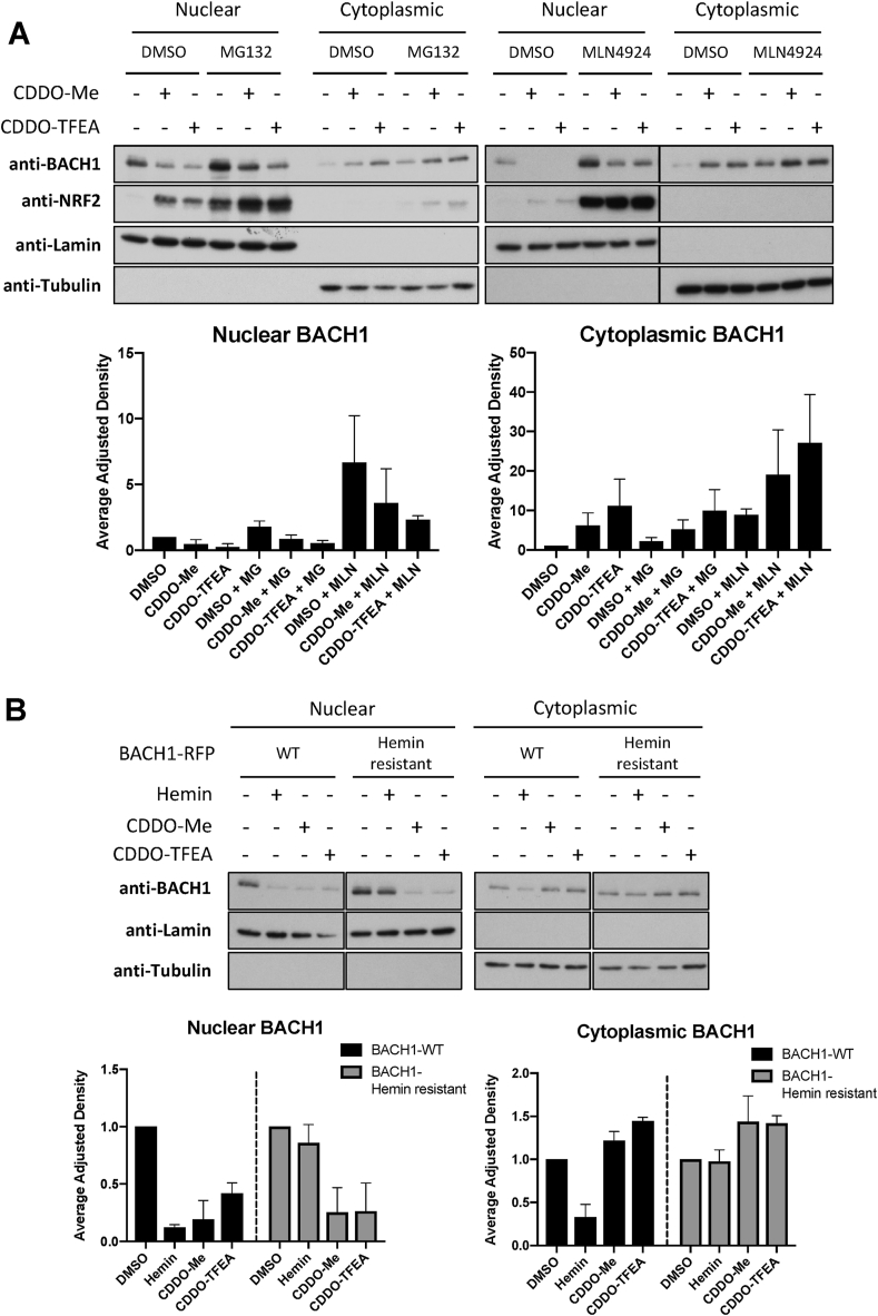
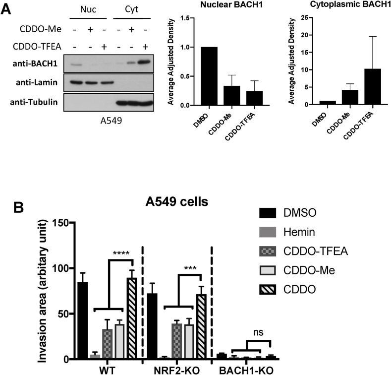
The transcription factor BACH1 is a potential therapeutic target for a variety of chronic conditions linked to oxidative stress and inflammation, as well as cancer metastasis. However, only a few BACH1 degraders/inhibitors have been described. BACH1 is a transcriptional repressor of heme oxygenase 1 (HMOX1), which is positively regulated by transcription factor NRF2 and is highly inducible by derivatives of the synthetic oleanane triterpenoid 2-cyano-3,12-dioxooleana-1,9(11)-dien-28-oic acid (CDDO). Most of the therapeutic activities of these compounds are due to their anti-inflammatory and antioxidant properties, which are widely attributed to their ability to activate NRF2. However, with such a broad range of action, these compounds have other molecular targets that have not been fully identified and could also be of importance for their therapeutic profile. Herein we identified BACH1 as a target of two CDDO-derivatives (CDDO-Me and CDDO-TFEA), but not of CDDO. While both CDDO and CDDO-derivatives activate NRF2 similarly, only CDDO-Me and CDDO-TFEA inhibit BACH1, which explains the much higher potency of these CDDO-derivatives as HMOX1 inducers compared with unmodified CDDO. Notably, we demonstrate that CDDO-Me and CDDO-TFEA inhibit BACH1 via a novel mechanism that reduces BACH1 nuclear levels while accumulating its cytoplasmic form. In an in vitro model, both CDDO-derivatives impaired lung cancer cell invasion in a BACH1-dependent and NRF2-independent manner, while CDDO was inactive. Altogether, our study identifies CDDO-Me and CDDO-TFEA as dual KEAP1/BACH1 inhibitors, providing a rationale for further therapeutic uses of these drugs.

Keywords: BACH1; CDDO; HMOX1; NRF2.


Conflict of interest statement

The authors declare the following financial interests/personal relationships which may be considered as potential competing interests: ADK is a member of the Scientific Advisory Board of Evgen Pharma, and a consultant for Aclipse Therapeutics.

Figures

Fig. 1
Fig. 1
CDDO-Me and CDDO-TFEA, but not CDDO, reduce BACH1 levels. (A) HaCaT cells were treated with either DMSO (0.1%, v/v), SFN (5 μM), CDDO (100 nM), TBE-31 (100 nM) or Hemin (10 μM) for 16 h. Cells were lysed and mRNA levels of HMOX1 and AKR1B10 were analysed by qRT-PCR, using HPRT1 as a housekeeping gene. ***P ≤ 0.001, ****P ≤ 0.0001. (B) As in A, but HaCaT cells were treated with either DMSO (0.1%, v/v) or increasing concentrations of CDDO or CDDO-Me. After 16 h cells were harvested and lysed and mRNA levels of HMOX1 and AKR1B10 were analysed by real-time qPCR. Data were normalised using HPRT1 as an internal control (n = 3) and are expressed relative to the DMSO treated sample. *P ≤ 0.05, **P ≤ 0.01, ***P ≤ 0.001. (C) HaCaT cells were treated with DMSO (0.1%, v/v) or increasing concentrations of CDDO or CDDO-Me. Five hours later, cells were harvested and lysed. Protein levels of NRF2, BACH1, HMOX1 and ACTIN were analysed by Western Blot. Left panel shows a representative blot and right panels show quantification of NRF2 and HMOX1 protein levels against the loading control. Data represent means ± SD (n = 3) and are expressed relative to the DMSO-treated samples. (D) HaCaT cells were treated with either DMSO (0.1%, v/v), CDDO-Me (100 nM) or CDDO-TFEA (100 nM) for 1 h, 3 h, 6 h or 16 h. Cells were harvested, lysed and analysed for the levels of the indicated proteins. Left panel is a representative blot; right panels are the quantification of BACH1 levels (n = 3). Data are expressed relative to the DMSO-treated sample at time 1 h, which is set to 1.
Fig. 2
Fig. 2
The differential effect of CDDO, CDDO-Me and CDDO-TFEA on HMOX1 expression is due to BACH1 inhibition. (A,B) HaCaT WT or NRF2-KO cells were treated with either DMSO (0.1%, v/v), CDDO (100 nM), CDDO-Me (100 nM) or CDDO-TFEA (100 nM) for 16 h. Samples were collected and mRNA levels of HMOX1 (A) and AKR1B10 (B) were analysed via real-time qPCR, using HPRT1 as an internal control. Data are expressed relative to the DMSO-treated samples in each cell line (DMSO in WT and NRF2-KO cells set to 1). *P ≤ 0.05, ***P ≤ 0.001, ****P ≤ 0.0001. (C) HK2 Control (WT) and NRF2-GOF cells were treated with DMSO, CDDO (100 nM), CDDO-Me (100 nM), CDDO-TFEA (100 nM) or Hemin (10 μM) for 16 h. HMOX1 mRNA levels were analysed using RT-qPCR and HPRT1 as a housekeeping gene. Data are expressed relative to the DMSO-treated samples in each cell line (DMSO in WT and NRF2-GOF cells set to 1). (D) HaCaT BACH1-KO and HaCaT NRF2/BACH1-KO cells were treated as in (A). Levels of HMOX1 were analysed by qRT-PCR as previously described. HMOX1 levels in the DMSO samples of each cell line were set to 1 and the rest of the data are expressed relative to their corresponding DMSO sample. *P ≤ 0.05, **P ≤ 0.01, ***P ≤ 0.001.
Fig. 3
Fig. 3
Both CDDO-Me and CDDO-TFEA reduce nuclear BACH1 while increasing cytoplasmic BACH1 levels. (A–B) HaCaT WT cells (A) and NRF2-KO cells (B) were treated with DMSO (0.1%, v/v) or CDDO-TFEA (100 nM) for 1 h, 3 h, 6 h or 16 h. Cells were harvested and nuclear and cytosolic fractions were isolated and analysed for the levels of the indicated proteins. Upper panel is a representative blot; lower panels are the quantification of BACH1 nuclear and cytoplasmic levels (n = 2). Data are expressed relative to the DMSO-treated samples for each time point (which were set to 1) and were normalised against their respective loading control. (G).
Fig. 4
Fig. 4
CDDO-Me and CDDO-TFEA affect BACH1 levels in a proteasome independent manner and have a mechanism of action different than hemin. (A) HaCaT cells were incubated with either DMSO (0.1%, v/v), MG132 (10 μM) or MLN4924 (2 μM) for 1 h. After that, either DMSO (−), CDDO-Me (100 nM) or CDDO-TFEA (100 nM) was added. Six hours later, cells were harvested and nuclear/cytoplasmic fractions were isolated and analysed for their levels of BACH1 and NRF2. Upper panel is a representative blot and lower panels are the quantifications of nuclear and cytoplasmic BACH1 levels normalised against their corresponding loading control. Data represent means ± SD (n = 3) and are expressed relative to the DMSO sample. (B) HaCaT BACH1-KO cells reconstituted with either BACH1-RFP-WT or BACH1-RFP-Hemin resistant mutant were treated with DMSO (−), Hemin (10 μM), CDDO-Me (100 nM) or CDDO-TFEA (100 nM) for 6 h. Cells were harvested and nuclear/cytoplasmic fractions were isolated and analysed for their levels of BACH1. Upper panel is a representative blot and lower panels are the quantifications of nuclear and cytoplasmic BACH1 levels normalised against their corresponding loading control. Data represent means ± SD (n = 3) and are expressed relative to their DMSO control (each cell line against their own DMSO, which was set as 1).
Fig. 5
Fig. 5
CDDO-Me and CDDO-TFEA reduce lung cancer cell invasion in a BACH1-dependent and NRF2-independent manner. (A) A549 cells were treated with DMSO (0.1%, v/v), CDDO-Me (100 nM) or CDDO-TFEA (100 nM). Six hours later cells were harvested and subcellular fractionation was performed. BACH1 protein levels were analysed via western blot. Panels on the left show a representative blot; panels on the right are the corresponding BACH1 nuclear and cytoplasmic quantifications, which were normalised against their internal control (i.e., LAMIN for nuclear and TUBULIN for cytoplasmic levels). Data represent means ± SD (n = 3) and are expressed relative to the DMSO-treated samples. (B) WT, NRF2-KO, or BACH1-KO A549 cells were treated with DMSO, CDDO (100 nM), CDDO-Me (100 nM), CDDO-TFEA (100 nM) or hemin (15 μM) for 6 h, followed by transwell invasion assays that were performed in presence of the inhibitors.

Similar articles

Cited by

References

    1. Wang Y., Porter W.W., Suh N., Honda T., Gribble G.W., Leesnitzer L.M., et al. A synthetic triterpenoid, 2-cyano-3,12-dioxooleana-1,9-dien-28-oic acid (CDDO), is a ligand for the peroxisome proliferator-activated receptor gamma. Mol. Endocrinol. 2000;14(10):1550–1556. - PubMed
    1. Konopleva M., Tsao T., Ruvolo P., Stiouf I., Estrov Z., Leysath C.E., et al. Novel triterpenoid CDDO-Me is a potent inducer of apoptosis and differentiation in acute myelogenous leukemia. Blood. 2002;99(1):326–335. - PubMed
    1. Liby K., Royce D.B., Williams C.R., Risingsong R., Yore M.M., Honda T., et al. The synthetic triterpenoids CDDO-methyl ester and CDDO-ethyl amide prevent lung cancer induced by vinyl carbamate in A/J mice. Cancer Res. 2007;67(6):2414–2419. - PubMed
    1. Sun K., Li M., Konopleva M., Konoplev S., Stephens L.C., Kornblau S.M., et al. The synthetic triterpenoid, CDDO, suppresses alloreactive T cell responses and reduces murine early acute graft-versus-host disease mortality. Biol. Blood Marrow Transplant. 2007;13(5):521–529. - PMC - PubMed
    1. Tran T.A., McCoy M.K., Sporn M.B., Tansey M.G. The synthetic triterpenoid CDDO-methyl ester modulates microglial activities, inhibits TNF production, and provides dopaminergic neuroprotection. J. Neuroinflammation. 2008;5:14. - PMC - PubMed

Publication types

MeSH terms