. 2022 Apr 8;3(4):262-268.e4.
doi: 10.1016/j.medj.2022.03.004. Epub 2022 Mar 17.

Reduced pathogenicity of the SARS-CoV-2 omicron variant in hamsters

Affiliations

Reduced pathogenicity of the SARS-CoV-2 omicron variant in hamsters

Katherine McMahan et al. Med. .

Abstract

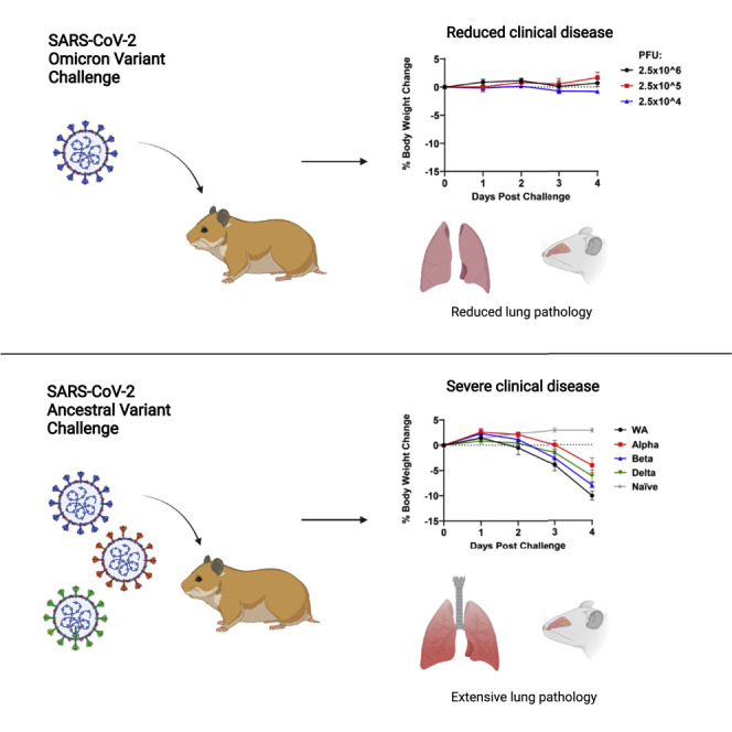
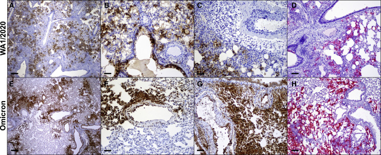
Background: The severe acute respiratory syndrome coronavirus 2 (SARS-CoV-2) Omicron (B.1.1.529) variant has proven to be highly transmissible and has outcompeted the Delta variant in many regions of the world. Early reports have also suggested that Omicron may result in less severe clinical disease in humans. Here, we show that Omicron is less pathogenic than prior SARS-CoV-2 variants in Syrian golden hamsters.

Methods: Hamsters were inoculated with either SARS-CoV-2 Omicron or other SARS-CoV-2 variants. Animals were followed for weight loss, and upper and lower respiratory tract tissues were assessed for viral loads and histopathology.

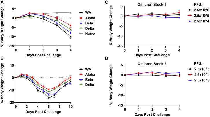
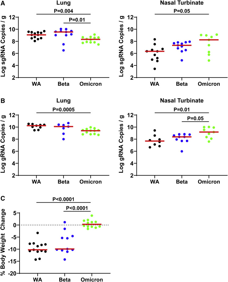
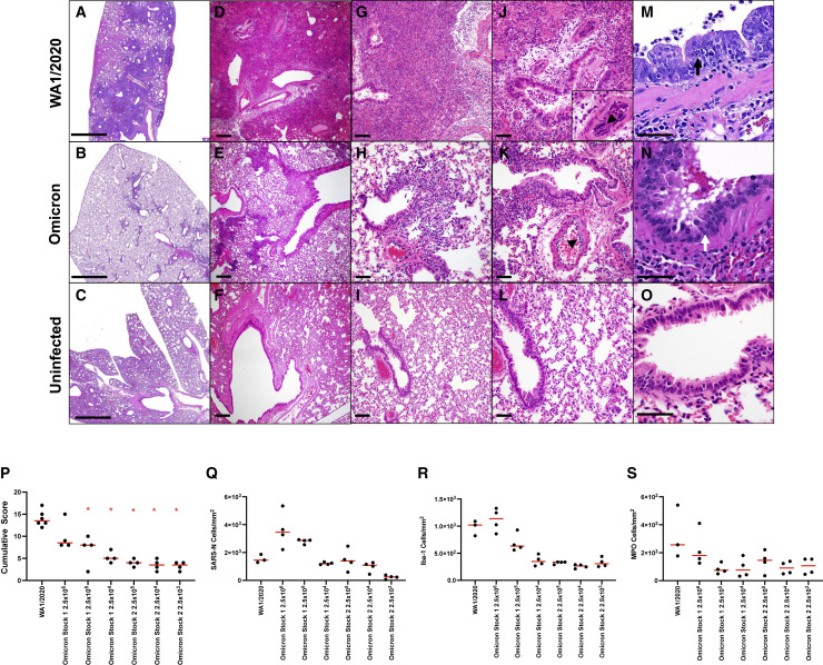
Findings: Infection of hamsters with the SARS-CoV-2 WA1/2020, Alpha, Beta, or Delta strains led to 4%-10% weight loss by day 4 and 10%-17% weight loss by day 6. In contrast, infection of hamsters with two different Omicron challenge stocks did not result in any detectable weight loss, even at high challenge doses. Omicron infection led to substantial viral replication in both the upper and lower respiratory tracts but demonstrated lower viral loads in lung parenchyma and reduced pulmonary pathology compared with WA1/2020 infection.

Conclusions: These data suggest that the SARS-CoV-2 Omicron variant may result in robust upper respiratory tract infection, but less severe lower respiratory tract clinical disease, compared with prior SARS-CoV-2 variants.

Funding: Funding for this study was provided by NIH grant CA260476, the Massachusetts Consortium for Pathogen Readiness, the Ragon Institute, and the Musk Foundation.

Keywords: COVID-19; SARS-CoV-2; hamster; omicron; pathogenicity.


Conflict of interest statement

The authors declare no competing interests.

Figures

None
Graphical abstract
Figure 1
Figure 1
Weight loss in hamsters infected with SARS-CoV-2 variants (A and B) Mean body-weight change following infection of hamsters with SARS-CoV-2 WA1/2020, Alpha, Beta, and Delta variants, along with weight change in uninfected control hamsters. (C and D) Mean body-weight change following infection of hamsters with SARS-CoV-2 Omicron stocks 1 and 2. Mean body-weight changes with standard errors are shown.
Figure 2
Figure 2
Tissue viral loads in hamsters on day 4 following SARS-CoV-2 infection (A) E subgenomic RNA (sgRNA) levels in lung and nasal turbinates following infection of hamsters with SARS-CoV-2 WA1/2020, Beta, and Omicron variants (limit of detection 100 viral copies/g tissue). (B) N genomic RNA (gRNA) levels in lung and nasal turbinates following infection of hamsters with SARS-CoV-2 WA1/2020, Beta, and Omicron variants (limit of detection 100 viral copies/g tissue). (C) Weight loss at time of necropsy of hamsters in (A) and (B). Data for WA1/2020 reflect N = 13 hamsters pooled from four different challenge experiments, data for Beta reflect N = 11 hamsters pooled from three different challenge experiments, and data for Omicron reflect N = 12 hamsters pooled from one experiment. Log sgRNA copies per gram tissue are shown. Medians (red bars) are depicted. p values represent two-sided Mann-Whitney tests.
Figure 3
Figure 3
Histopathology in hamsters on day 4 following SARS-CoV-2 infection Lung tissue from hamsters infected with 5 × 104 PFU SARS-CoV-2 WA1/2020 (top) and 2.5 × 105 PFU Omicron (middle) compared with uninfected hamsters (bottom) was stained with H&E. (A–C) Low power-representative images of lung. There are multifocal to locally extensive areas of interstitial inflammation and consolidation associated with bronchioles in WA1/2020-infected animals that are reduced in Omicron-infected animals. (D–L) Medium power-representative images of bronchioles and lumina showing presence of intraluminal necrotic epithelium in both WA1/2020- and Omicron-infected animals (J–L) and perivascular edema and marginating inflammatory cells along the endothelium of medium-sized arterioles (arrowheads). (M–O) High magnification images of bronchiolar epithelium showing cellular atypia, hypertrophy, and loss of basal nuclear polarity in degenerative bronchiolar epithelium that is more pronounced in WA1/2020-infected hamsters compared with Omicron-infected hamsters. (P) Cumulative pathology scoring of (1) airways (bronchi, bronchioles), (2) interstitium, (3) alveoli, (4) vessels, (5) edema, and (6) regeneration. Each feature received a score of 0–3 with a maximum possible score of 18 per animal. (Q–S) Quantitative image analysis of immunohistochemistry using HALO (Indicalabs)-optimized algorithms to enumerate (Q) SARS-N-positive (R), Iba-1-positive (macrophages), and (S) myeloperixade-positive (neutrophils) cells per unit area. ∗p = 0.0048, two-tailed Mann-Whitney test. Scale bars: 2 mm (A–C), 200 μm (D–F), 100 μm (G–I), and 50 μM (J–O).
Figure 4
Figure 4
SARS-CoV-2 Omicron variant distribution in lung (A–H) Immunohistochemistry for nucleocapsid protein (brown) and RNAscope in situ hybridization for viral RNA (red) in hamsters infected with SARS-CoV2 WA1/2020 (A–D) and Omicron (E–H). Scale bars: 200 μm (A and E) and 50 μm (B–D and F–H).

References

    1. Cele S., Jackson L., Khoury D.S., Khan K., Moyo-Gwete T., Tegally H., San J.E., Cromer D., Scheepers C., Amoako D.G., et al. Omicron extensively but incompletely escapes Pfizer BNT162b2 neutralization. Nature. 2021;602:654–656. doi: 10.1038/s41586-021-04387-1. - DOI - PMC - PubMed
    1. Tostanoski L.H., Wegmann F., Martinot A.J., Loos C., McMahan K., Mercado N.B., Yu J., Chan C.N., Bondoc S., Starke C.E., et al. Ad26 vaccine protects against SARS-CoV-2 severe clinical disease in hamsters. Nat. Med. 2020;26:1694–1700. doi: 10.1038/s41591-020-1070-6. - DOI - PMC - PubMed
    1. Chan J.F., Zhang A.J., Yuan S., Poon V.K., Chan C.C., Lee A.C., Chan W.M., Fan Z., Tsoi H.W., Wen L., et al. Simulation of the clinical and pathological manifestations of coronavirus disease 2019 (COVID-19) in golden syrian hamster model: implications for disease pathogenesis and transmissibility. Clin. Infect. Dis. 2020;71:2428–2446. doi: 10.1093/cid/ciaa325. - DOI - PMC - PubMed
    1. Tostanoski L.H., Yu J., Mercado N.B., McMahan K., Jacob-Dolan C., Martinot A.J., Piedra-Mora C., Anioke T., Chang A., Giffin V.M., et al. Immunity elicited by natural infection or Ad26.COV2.S vaccination protects hamsters against SARS-CoV-2 variants of concern. Sci. Transl. Med. 2021;13:eabj3789. doi: 10.1126/scitranslmed.abj3789. - DOI - PMC - PubMed
    1. Dagotto G., Mercado N.B., Martinez D.R., Hou Y.J., Nkolola J.P., Carnahan R.H., Crowe J.E., Jr., Baric R.S., Barouch D.H. Comparison of subgenomic and total RNA in SARS-CoV-2 challenged rhesus macaques. J. Virol. 2021;95:e02370–e02420. doi: 10.1128/JVI.02370-20. - DOI - PMC - PubMed

Publication types

Supplementary concepts