Review
. 2022 Mar 1;25(4):103994.
doi: 10.1016/j.isci.2022.103994. eCollection 2022 Apr 15.

Cross-kingdom microbial interactions in dental implant-related infections: is Candida albicans a new villain?

Affiliations
Review

Cross-kingdom microbial interactions in dental implant-related infections: is Candida albicans a new villain?

João G S Souza et al. iScience. .

Abstract

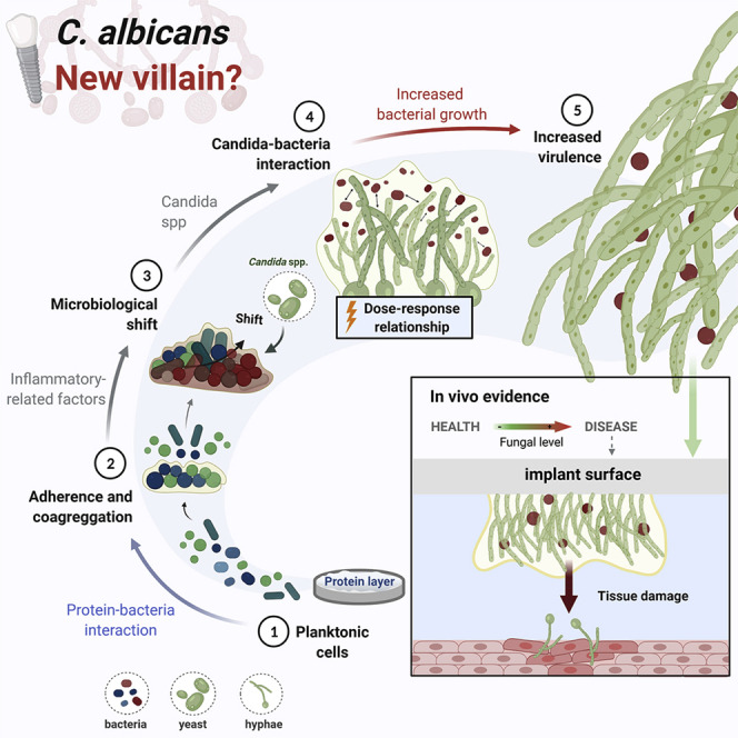
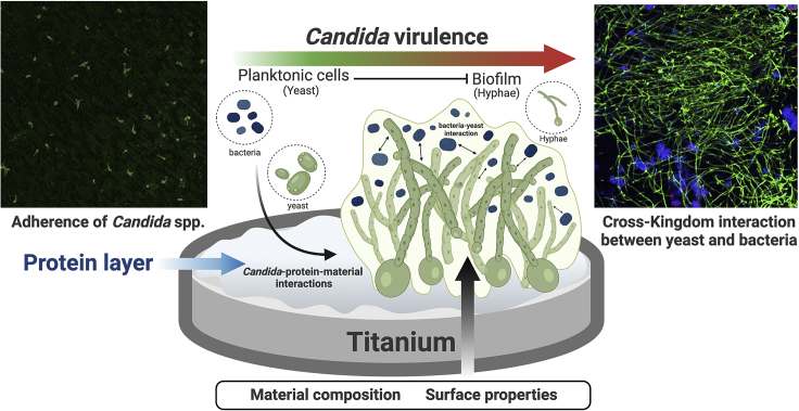
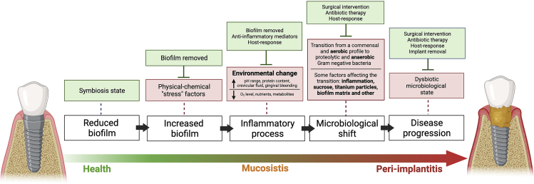
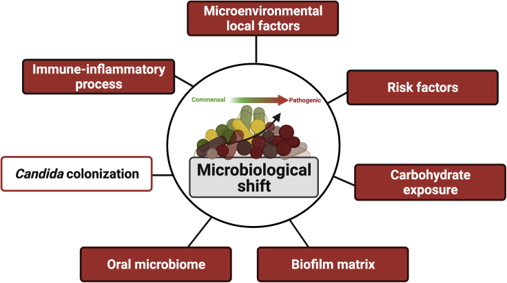
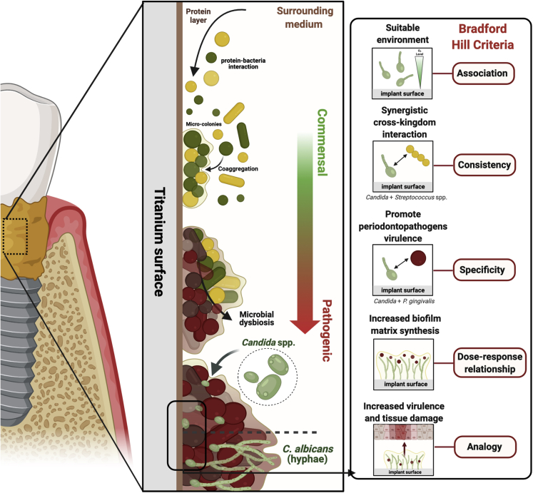
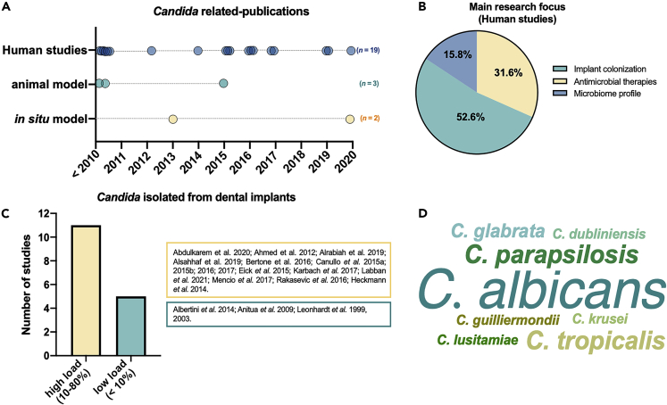
Candida albicans, an oral fungal opportunistic pathogen, has shown the ability to colonize implant surfaces and has been frequently isolated from biofilms associated with dental implant-related infections, possibly due to its synergistic interactions with certain oral bacteria. Moreover, evidence suggests that this cross-kingdom interaction on implant can encourage bacterial growth, leading to increased fungal virulence and mucosal damage. However, the role of Candida in implant-related infections has been overlooked and not widely explored or even considered by most microbiological analyses and therapeutic approaches. Thus, we summarized the scientific evidence regarding the ability of C. albicans to colonize implant surfaces, interact in implant-related polymicrobial biofilms, and its possible role in peri-implant infections as far as biologic plausibility. Next, a systematic review of preclinical and clinical studies was conducted to identify the relevance and the gap in the existing literature regarding the role of C. albicans in the pathogenesis of peri-implant infections.

Keywords: Mycology; microbiofilms.


Conflict of interest statement

The authors declare no competing interests.

Figures

None
Graphical abstract
Figure 1
Figure 1
Relationship between material/substrate type and Candida biofilm development Representative microscopy of acrylic (Poly(methyl methacrylate) - PMMA) and titanium surface topography evaluated by white light 3D profilometry (left side) Scanning electron microscopy (SEM) images (right side) at 1.5 h (adhesion) and 48 h (maturation) of C. albicans biofilm formed on PMMA (top) and titanium (bottom). White arrows indicate the presence of hyphae, which were prevalent in both materials at 48 h. Overall, the similar surface roughness of both materials and the presence of environment fluids (saliva and blood plasma) culminated in no difference for Candida adhesion and growth for the two tested substrates. Reprinted (adapted) from ref (Cavalcanti et al., 2016a; 2016b); Copyright (2016), with permission from Elsevier (License number: 5117881126618).
Figure 2
Figure 2
Schematic representation of Candida spp. attachment and accumulation on Ti surface After implantation, the Ti-based implant surface is immediately coated by the protein layer. Consequently, microbial-material interactions promote Candida adhesion (confocal images, left side; scale: 50 μm/time: 2 h - stained by immuno-FISH green). Moreover, fungal biofilm growth is modulated by surface and microorganism properties and environmental conditions, leading to the transition of Candida morphology from yeast to hyphae in single or multi-species biofilms. The cross-kingdom interaction between Candida and Streptococcus on implant surface may promote biofilm growth. It is also possible to observe in the confocal image (right side) the mixed biofilm stained by immuno-FISH of Candida albicans (green) and Streptococcus oralis stained by Streptococcus-specific probe conjugated to Alexa 405 (blue) growing on Ti surface (scale: 50 μm/time: 72h). Reprinted (adapted) from refs (Souza et al., 2020b; 2020d). Created with BioRender.com (license number: FH22TBHYN9).
Figure 3
Figure 3
Schematic representation of the “ecological plaque hypothesis” concerning peri-implant disease, adapted from Marsh et al. (2011), Rosier et al. (2018), and Souza et al., 2020a, , , , , Increased biofilm accumulation on implant surface triggers an inflammatory process that changes the environment leading to microbiological shift and disease progression, as shown by red boxes. Other factors can also favor the microbiological shift on biofilms growing on titanium surfaces, such as carbohydrate (sucrose exposure). However, some factors can control biofilm accumulation and inflammatory response, shown in green boxes, such as surgical and antimicrobial intervention and host response.
Figure 4
Figure 4
Different factors directly affect microbiological shift on implant-related biofilms from a commensal to a pathogenic profile, such as extracellular biofilm matrix, inflammatory process, and carbohydrate exposure Because Candida colonization promotes biofilm accumulation and virulence factors, this opportunistic pathogen should be considered an additional factor leading to microbiological shift on implant-related biofilms, which must be tested experimentally. Created with BioRender.com (license number: ZV22TBI7ME).
Figure 5
Figure 5
Schematic representation of the role of Candida to promote biofilm accumulation and virulence with expected higher tissue damage on peri-implantitis The biological plausibility hypothesis of Candida’|'s role on peri-implantitis was considered based on current evidence and Bradford Hill criteria. (A–E) Implant surface and surrounding microenvironment (i.e., low oxygen level) seem a suitable site for Candida colonization, mainly after protein pellicle adsorption; (B) Candida interactions with bacteria highly associated with peri-implantitis, such as P. gingivalis, promoting bacterial growth, virulence, and the ability to invade host cells; (C) Cross-kingdom and synergistic interaction with Streptococcus species, group highly found in healthy and disease implant sites, forming a hyper-virulent biofilm with increased bacterial biomass, upregulating hyphae-related genes, exacerbated inflammatory response, and tissue damage; (D) Candida increases the extracellular polymers synthesis by bacterial species, enhancing biofilm matrix which has several advantages for microbial accumulation, leading to microbial dysbiosis and increased virulence; therefore, high Candida count is expected to lead to increased biofilm matrix synthesis; (E) The effect on bacteria growth and biofilm virulence lead to increased tissue damage. Created with BioRender.com (license number: CA22TEEYEL).
Figure 6
Figure 6
Results of the systematic review to identify in vivo studies evaluating the presence or level of Candida on the implant surface (A) Twenty-four in vivo studies were included among animal, in situ, and human models. (B) The main research focus of included studies. (C) The number of studies describing high (10%–80%) or low (<10%) Candida load on the implant surface. (D) Word cloud graph of Candida species found on implant surface according to the number of studies.

Similar articles

Cited by

References

    1. Ahmed A., Chambers M.S., Goldschmidt M.C., Habib A., Lei X., Jacob R.F. Association between microbial flora and tissue abnormality around dental implants penetrating the skin in reconstructed oral cancer patients. Int. J. Oral Maxill. Implants. 2012;27:684–694. - PubMed
    1. Alqahtani F. Role of oral yeasts in the etiopathogenesis of peri-implantitis: an evidence-based literature review of clinical studies. Arch. Oral Biol. 2020;111:104650. doi: 10.1016/j.archoralbio.2020.104650. - DOI - PubMed
    1. Alrabiah M., Alshagroud R.S., Alsahhaf A., Almojaly S.A., Abduljabbar T., Javed F. Presence of Candida species in the subgingival oral biofilm of patients with peri-implantitis. Clin. Implant Dent Relat. Res. 2019;21:781–785. doi: 10.1111/cid.12760. - DOI - PubMed
    1. Alsahhaf A., Al-Aali K.A., Alshagroud R.S., Alshiddi I.F., Alrahlah A., Abduljabbar T., Javed F., Vohra F. Comparison of yeast species in the subgingival oral biofilm of individuals with type 2 diabetes and peri-implantitis and individuals with peri-implantitis without diabetes. J. Periodontol. 2019;90:1383–1389. doi: 10.1002/JPER.19-0091. - DOI - PubMed
    1. Andes D., Nett J., Oschel P., Albrecht R., Marchillo K., Pitula A. Development and characterization of an in vivo central venous catheter Candida albicans biofilm model. Infect. Immun. 2004;72:6023–6031. doi: 10.1128/IAI.72.10.6023-6031.2004. - DOI - PMC - PubMed

LinkOut - more resources