

A Robust and Highly Efficient Approach for Isolation of Mesenchymal Stem Cells From Wharton's Jelly for Tissue Repair
- PMID: 35313748
- PMCID: PMC8943591
- DOI: 10.1177/09636897221084354
A Robust and Highly Efficient Approach for Isolation of Mesenchymal Stem Cells From Wharton's Jelly for Tissue Repair
Abstract
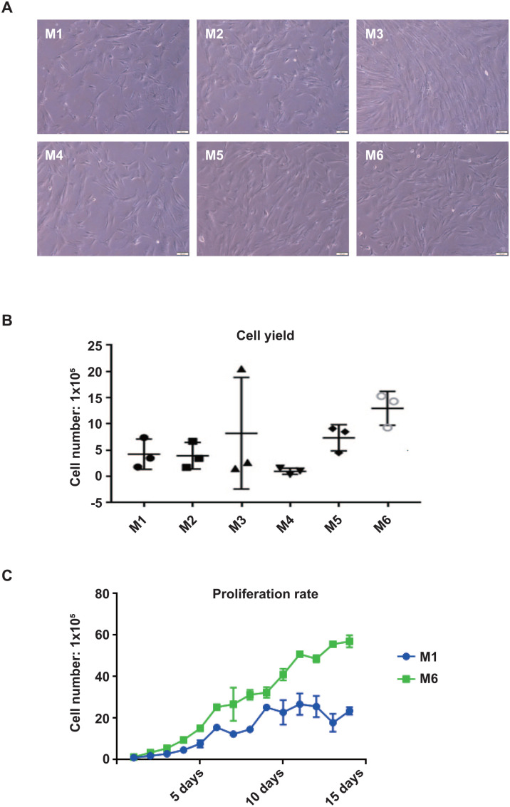
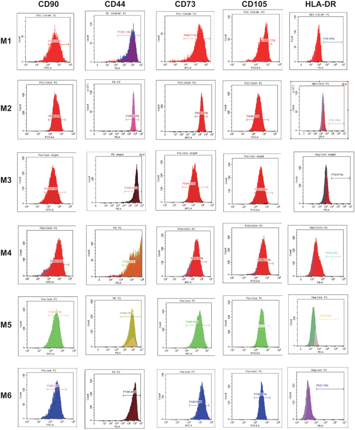
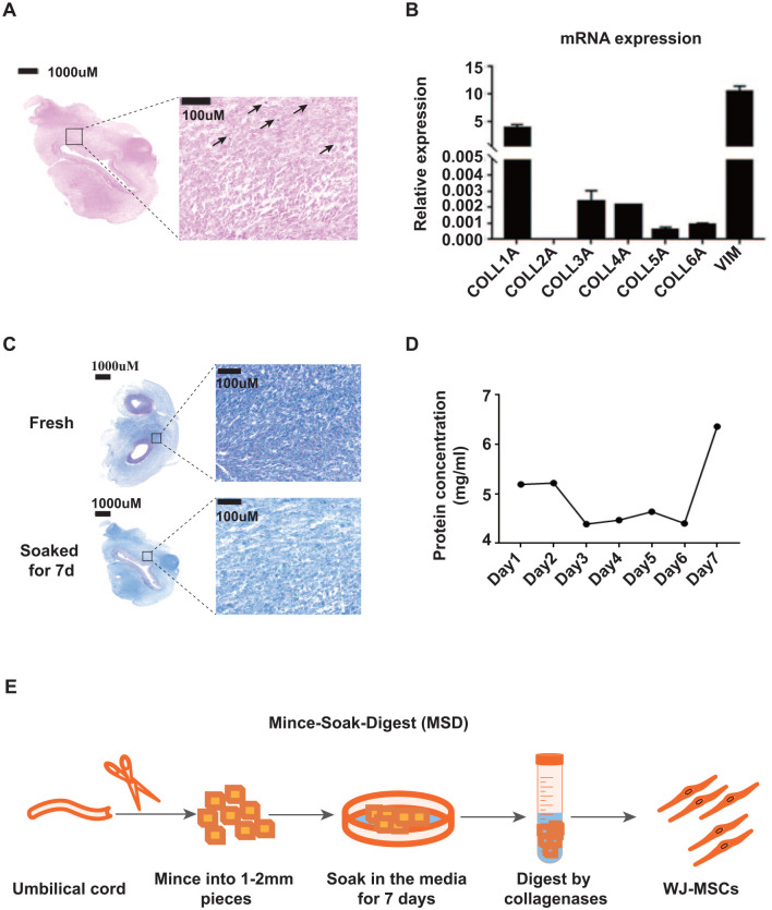
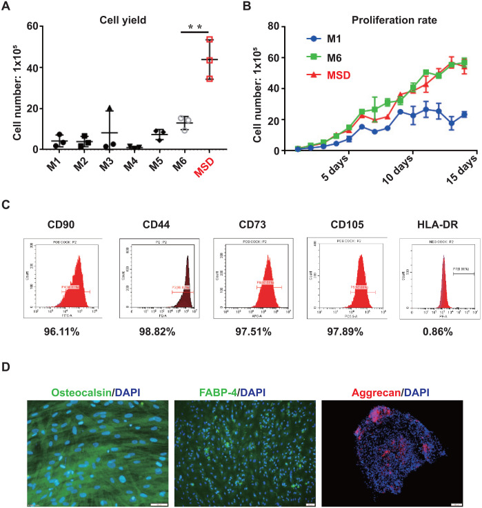
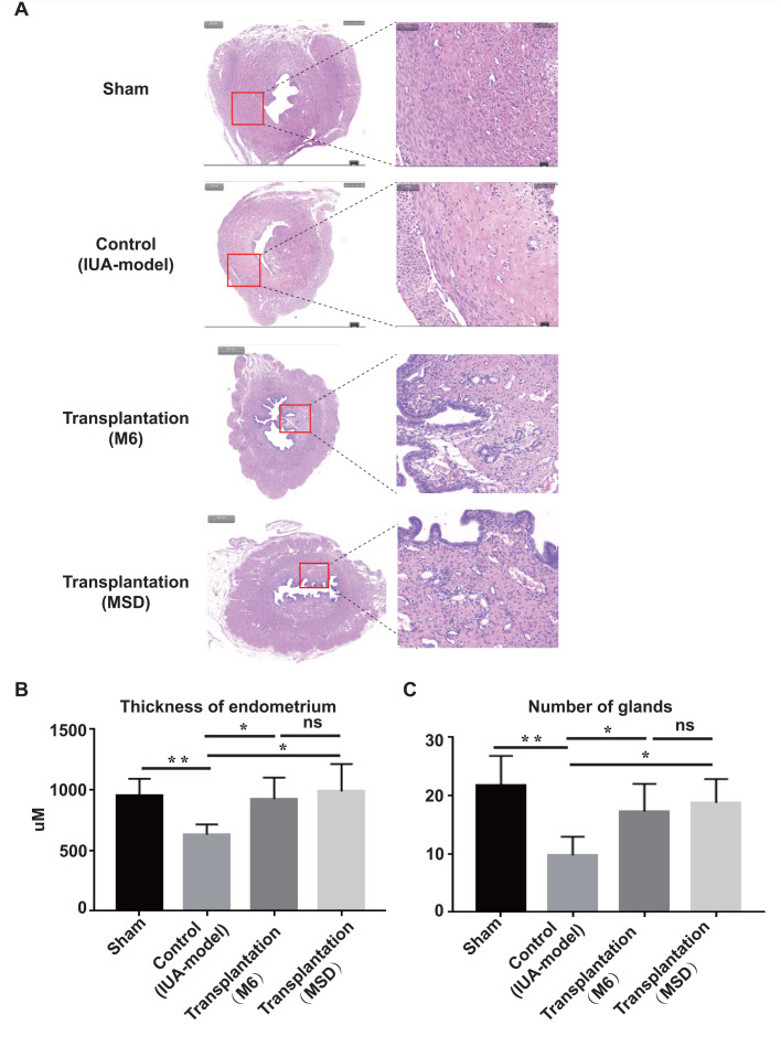
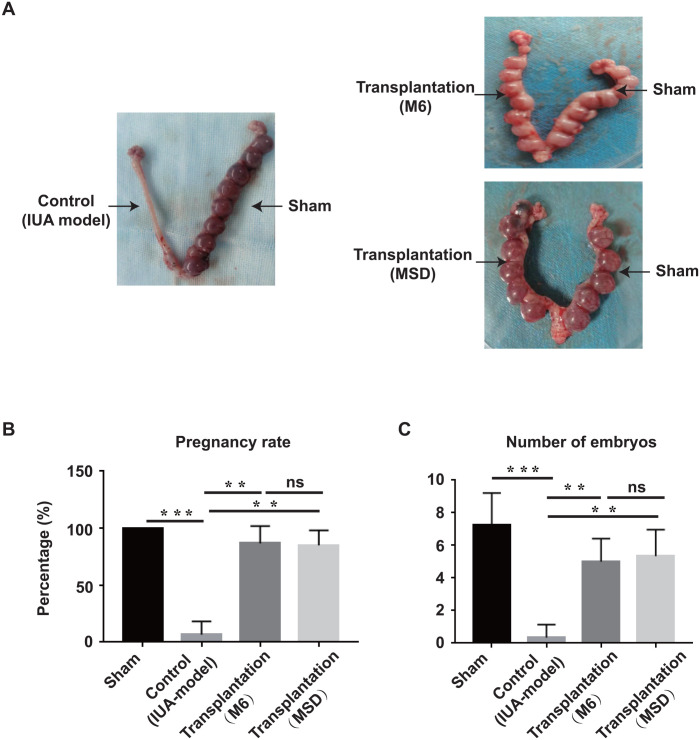
Mesenchymal stem cells derived from umbilical cord Wharton's Jelly (WJ-MSCs) are emerging as promising therapeutics for a variety of diseases due to their ability of regeneration and immunomodulation, and their non-tumorigenic and non-immunogenic properties. Although multiple protocols have been developed for WJ-MSC isolation, insufficient cell numbers, heterogeneous cell population, and variations in procedures between different laboratories impede further clinical applications. Here, we compared six widely used WJ-MSC isolation methods regarding cell morphology, yield, purity, proliferation rate, and differentiation potential. Based on these analyses, we identified that the inefficiency of the extracellular matrix digestion results in low cell yield. Thus, we developed a new method called "Mince-Soak-Digest (MSD)" to isolate MSCs from WJ by incorporating a soaking step to facilitate the digestion of the extracellular matrix and release of the cells. Our newly developed method generates significantly higher cell yield (4- to 10-fold higher) than six widely used methods that we tested with high purity and consistency. Importantly, by transplantation of WJ-MSCs to the rat uterus, we repair the endometrial injury and restore the fertility of the rats. In conclusion, our results provide a robust and highly efficient approach for the isolation of WJ-MSCs to restore injured tissue. The higher efficiency of MSD assures the abundance of WJ-MSCs for clinical applications. Furthermore, the reliability of MSD contributes to the standardization of WJ-MSC isolation, which eliminates the discrepancies due to isolation procedures, thus facilitating the evaluation of the efficacy of WJ-MSCs across various human clinical applications.
Keywords: Wharton’s jelly; intrauterine adhesions; mesenchymal stem cells; tissue repair; transplantation.
Conflict of interest statement
Figures
Similar articles
-
Differential expression of cell cycle and WNT pathway-related genes accounts for differences in the growth and differentiation potential of Wharton's jelly and bone marrow-derived mesenchymal stem cells.Stem Cell Res Ther. 2017 Apr 26;8(1):102. doi: 10.1186/s13287-017-0555-9. Stem Cell Res Ther. 2017. PMID: 28446235 Free PMC article.
-
Differentiation of human umbilical cord Wharton's jelly-derived mesenchymal stem cells into endometrial cells.Stem Cell Res Ther. 2017 Nov 2;8(1):246. doi: 10.1186/s13287-017-0700-5. Stem Cell Res Ther. 2017. PMID: 29096715 Free PMC article.
-
A GMP-compliant manufacturing method for Wharton's jelly-derived mesenchymal stromal cells.Stem Cell Res Ther. 2024 May 3;15(1):131. doi: 10.1186/s13287-024-03725-0. Stem Cell Res Ther. 2024. PMID: 38702793 Free PMC article.
-
Characteristics and clinical applications of Wharton's jelly-derived mesenchymal stromal cells.Curr Res Transl Med. 2020 Jan;68(1):5-16. doi: 10.1016/j.retram.2019.09.001. Epub 2019 Sep 19. Curr Res Transl Med. 2020. PMID: 31543433 Review.
-
Regenerative potential of Wharton's jelly-derived mesenchymal stem cells: A new horizon of stem cell therapy.J Cell Physiol. 2020 Dec;235(12):9230-9240. doi: 10.1002/jcp.29810. Epub 2020 Jun 18. J Cell Physiol. 2020. PMID: 32557631 Review.
Cited by
-
The Role of Mesenchymal Stem Cells in Modulating the Breast Cancer Microenvironment.Cell Transplant. 2023 Jan-Dec;32:9636897231220073. doi: 10.1177/09636897231220073. Cell Transplant. 2023. PMID: 38135917 Free PMC article. Review.
-
Human Umbilical Cord-Based Therapeutics: Stem Cells and Blood Derivatives for Female Reproductive Medicine.Int J Mol Sci. 2022 Dec 14;23(24):15942. doi: 10.3390/ijms232415942. Int J Mol Sci. 2022. PMID: 36555583 Free PMC article. Review.
-
Evolution of biotechnological advances and regenerative therapies for endometrial disorders: a systematic review.Hum Reprod Update. 2024 Oct 1;30(5):584-613. doi: 10.1093/humupd/dmae013. Hum Reprod Update. 2024. PMID: 38796750 Free PMC article.
-
Human embryonic stem cell-derived immunity-and-matrix-regulatory cells promote endometrial repair and fertility restoration in IUA rats.Stem Cell Res Ther. 2025 Apr 23;16(1):204. doi: 10.1186/s13287-025-04298-2. Stem Cell Res Ther. 2025. PMID: 40269948 Free PMC article.
-
Long-term therapeutic effects of allogeneic mesenchymal stem cell transplantation for intrauterine adhesions.Stem Cell Res Ther. 2024 Dec 23;15(1):499. doi: 10.1186/s13287-024-04100-9. Stem Cell Res Ther. 2024. PMID: 39716301 Free PMC article.
References
Publication types
MeSH terms
LinkOut - more resources
Full Text Sources
