Case Reports
. 2022 Jan-Dec:10:23247096221086453.
doi: 10.1177/23247096221086453.

Pulmonary Tumor Embolism and Pulmonary Tumor Thrombotic Microangiopathy Causing Rapidly Progressive Respiratory Failure: A Case Series

Affiliations
Case Reports

Pulmonary Tumor Embolism and Pulmonary Tumor Thrombotic Microangiopathy Causing Rapidly Progressive Respiratory Failure: A Case Series

Kartikeya Rajdev et al. J Investig Med High Impact Case Rep. 2022 Jan-Dec.

Abstract

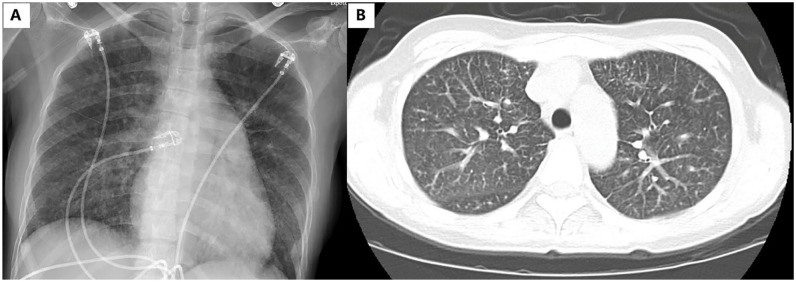
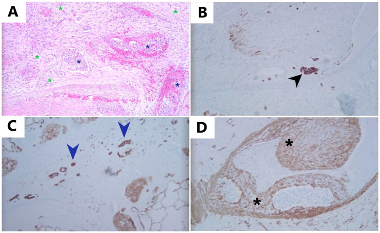
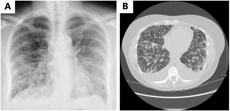
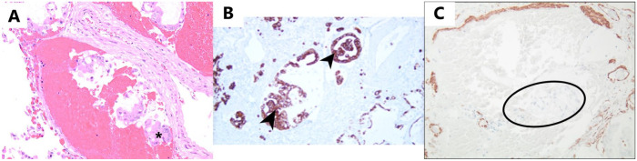
Pulmonary tumor embolism (PTE) and pulmonary tumor thrombotic microangiopathy (PTTM) are rare etiologies for rapidly progressive dyspnea in the setting of undiagnosed metastatic cancer. They occur most frequently in association with adenocarcinomas, with PTE being most frequently associated with hepatocellular carcinoma and PTTM being most commonly reported with gastric adenocarcinoma. Pulmonary tumor embolism and PTTM appear to be a disease spectrum where PTTM represents an advanced form of PTE. Pulmonary tumor embolism and PTTM are mostly identified postmortem during autopsy as the antemortem diagnosis remains a clinical challenge due to the rapidly progressive nature of these rare diseases. We report 2 cases of rapidly progressive respiratory failure leading to death, due to tumoral pulmonary hypertension resulting from PTE and PTTM, diagnosed postmortem. Both of the patients were middle-aged females, nonsmokers, and had a gastrointestinal source of their primary malignancy.

Keywords: PTE; PTTM; autopsy; cor pulmonale; metastatic cancer; pulmonary tumor embolism; pulmonary tumor thrombotic microangiopathy; respiratory failure; tumoral pulmonary hypertension.


Conflict of interest statement

Declaration of Conflicting Interests: The author(s) declared no potential conflicts of interest with respect to the research, authorship, and/or publication of this article.

Figures

Figure 1.
Figure 1.
(A) Chest radiograph revealing mild diffuse interstitial prominence and (B) CT angiography of chest revealing mild centrilobular nodular pattern and interlobular septal thickening. Abbreviation: CT, computed tomography.
Figure 2.
Figure 2.
Pulmonary tumor thrombotic microangiopathy: (A): H&E, 10×: H&E-stained slide showing fibrocellular intimal proliferation with spindle-shaped cells (green asterisks) surrounding the pulmonary tumor emboli (blue asterisks) and completely filling the lumen of this vessel, (B) Immunostaining for CK-7, 10×: CK-7 highlights tumor cells (black arrowhead) within the lumen of muscular arteries, adjacent to intimal proliferation, (C) Immunostaining for CK-7, 10×: CK-7-positive tumor cells (blue arrowheads) present in lymphatics, and (D) Immunostaining for SMA, 10×: smooth muscle actin (SMA) highlights proliferation of smooth muscle cells within tunica intima (intimal proliferation) (black asterisks).
Figure 3.
Figure 3.
(A) Chest radiograph at initial presentation revealing diffuse reticular opacities and tiny bilateral pleural effusions and (B) CT scan of chest without contrast revealing bilateral pleural effusions with diffuse interstitial septal thickening and scattered ground-glass opacities. Abbreviation: CT, computed tomography.
Figure 4.
Figure 4.
Pulmonary tumor embolism: (A) H&E 10×: H&E-stained slide showing tumor cells (asterisk) within the lumen of a muscular artery, causing venous congestion without intimal proliferation, (B) Immunostaining for CK-7, 10×: CK-7 positive tumor cells (black arrowheads) in the lumen of blood vessels, and (C) Immunostaining for SMA, 10×: tumor cells (outlined in black) within the lumen of the artery without causing intimal proliferation. SMA staining is confined to the original wall of the artery and not extending into the lumen. Abbreviation: SMA, smooth muscle actin.

References

    1. Uruga H, Fujii T, Kurosaki A, et al.. Pulmonary tumor thrombotic microangiopathy: a clinical analysis of 30 autopsy cases. Intern Med. 2013;52(12):1317-1323. - PubMed
    1. Price LC, Seckl MJ, Dorfmüller P, et al.. Tumoral pulmonary hypertension. Eur Respir Rev. 2019;28(151):180065. - PMC - PubMed
    1. Schmidt MB. Die Verbreitungswege der Karzinome und die Beziehung generalisierter Sarcome zu den leukaemischen Neubildungen. Fischer; 1903.
    1. Bassiri AG, Haghighi B, Doyle RL, et al.. Pulmonary tumor embolism. Am J Respir Crit Care Med. 1997;155(6):2089-2095. - PubMed
    1. Brill IC, Robertson TD. SUBACUTE COR PULMONALE. Arch Intern Med (Chic). 1937;60(6):1043-1057.

Publication types

MeSH terms