. 2022 Mar 21;32(1):4.
doi: 10.1186/s12610-022-00154-y.

A rare case of penile schwannomatosis presenting with painful nocturnal penile tumescence

Affiliations

A rare case of penile schwannomatosis presenting with painful nocturnal penile tumescence

Chan Ming Tow et al. Basic Clin Androl. .

Abstract

Background: Penile schwannoma is a rare tumor. They commonly present as an asymptomatic, painless and slow growing mass. Other presentations include sexual dysfunction, most commonly dyspareunia, followed by erectile dysfunction, abnormal penile curvature or pain with ejaculation.

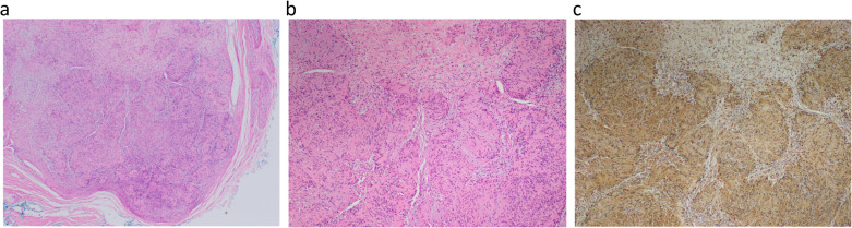
Case presentation: A 26-year-old male presented atypically with painful nocturnal penile tumescence, along with multiple nodules over the dorsal penis. Excision of multiple penile tumors under general anaesthesia was performed and histopathologic examination revealed benign schwannoma.

Conclusion: Our hypothesis is that the schwannoma lies along the axis of the dorsal penile nerve, and compression of this nerve occurs during his erection causing pain. However, there are limited presentations of painful erections in penile schwannomas, and we hope that future studies can help confirm this theory.

Abstraite: CONTEXTE: Le schwannome pénien est. une tumeur rare. Il se présente généralement comme une masse asymptomatique, indolore et à croissance lente. D’autres présentations incluent la dysfonction sexuelle, le plus souvent la dyspareunie, suivie de la dysfonction érectile, de la courbure anormale du pénis ou de la douleur à l’éjaculation. PRéSENTATION DU CAS: Un homme de 26 ans s’est. présenté de façon atypique avec une tumescence pénienne nocturne douloureuse, ainsi que de multiples nodules sur la face dorsale du pénis. L’excision de plusieurs tumeurs du pénis a été réalisée sous anesthésie générale et un examen histopathologique a révélé un schwannome bénin.

Conclusion: Notre hypothèse est. que le schwannome se trouve localisé le long de l’axe du nerf pénien dorsal, et que la compression de ce nerf se produit pendant l’érection, constituant la source des douleurs. Cependant, il existe peu de présentations d’érections douloureuses dans les schwannomes péniens, et nous espérons que de futures études pourront aider à confirmer cette théorie.

Keywords: Painful tumescence; Penile; Schwannoma; Sexual dysfunction.


Conflict of interest statement

The authors declare that they have no competing interests.

Figures

Fig. 1
Fig. 1
Ultrasound image (longitudinal view) of penile nodules. Ultrasound image (longitudinal view) of penile nodules demonstrates three well-circumscribed, hypoechoic nodules adjacent to the dorsal aspect of the corpus cavernosa
Fig. 2
Fig. 2
Ultrasound image (longitudinal view) of penile nodules with power doppler interrogation. Ultrasound image (longitudinal view) with power doppler interrogation reveals the presence of internal vascularity within the nodule
Fig. 3
Fig. 3
Coronal T2-weighted magnetic resonance image (MRI) of penile nodules. Coronal T2-weighted MR image shows three well-defined T2W hyperintense nodules (arrows) within the penis. These are superficial to the tunica albuginea (arrowhead)
Fig. 4
Fig. 4
Axial T2-weighted MRI of penis and nodule. Axial T2-weighted MRI of hyperintense nodule (arrow) is seen within the right corpus cavernosa
Fig. 5
Fig. 5
Sagittal T1-weighted post contrast MRI of penile nodules. Sagittal T1-weighted post contrast MRI of penile nodules displaying low signal intensity (arrows)
Fig. 6
Fig. 6
Coronal T1-weighted post contrast MRI of penile nodules. Coronal T1-weighted post contrast MRI showing penile nodules of low signal intensity with homogenous and avid post contrast enhancement (arrows)
Fig. 7
Fig. 7
Intra-operative image of excision of penile nodule. Intra-operative image of excision of penile nodule where the penis has been degloved to its base and the layers dissected down to Buck’s fascia. A superficial tumor measuring 1 cm in diameter can be seen
Fig. 8
Fig. 8
a, b, c Histologic section from excision biopsies. a Well-circumscribed and thinly encapsulated tumour nodule (Hematoxylin and Eosin stain, 20x magnification). b Cellular Antoni A areas with short fascicles of Schwann cells and less cellular Antoni B areas with myxoid stroma (Hematoxylin and Eosin stain, 200x magnification). c Diffuse and strong S100 expression in the tumour cells (S100 immunohistochemistry stain, 200x magnification)

References

    1. Loeser A, Katzenberger T, Meuller JG, Riedmiller H, Gerharz EW. Solitary schwannoma of the glans penis. Urol J. 2007;70(5):1007.e5-6. - PubMed
    1. Marshall J, Lin E, Dogra V, Davis R. Schwannoma of the penis: Preservation of the neurovascular bundle. Urol J. 2007;70(2):373.e1-3. - PubMed
    1. Schulz A, Büttner R, Hagel C, Baader S, Kluwe L, Salamon J, et al. The importance of nerve microenvironment for schwannoma development. Acta Neuropathol. 2016;132(2):289–307. doi: 10.1007/s00401-016-1583-8. - DOI - PMC - PubMed
    1. Hanemann C, Evans D. News on the genetics, epidemiology, medical care and translational research of schwannomas. J Neurol. 2006;253(12):1533–1541. doi: 10.1007/s00415-006-0347-0. - DOI - PubMed
    1. Parra CA. Salitary neurinomas of the glans penis. Dermatologica. 1968;137(3):150–155. doi: 10.1159/000254044. - DOI - PubMed

LinkOut - more resources