

Assessment of radial artery atherosclerosis in acute coronary syndrome patients: an in vivo study using optical coherence tomography
- PMID: 35313827
- PMCID: PMC8939080
- DOI: 10.1186/s12872-022-02561-5
Assessment of radial artery atherosclerosis in acute coronary syndrome patients: an in vivo study using optical coherence tomography
Abstract
Background: Radial artery (RA) atherosclerosis in acute coronary syndrome (ACS) patients has not been systematically observed in vivo. The study aims to characterize plaque morphology and intimal hyperplasia of the RA in patients with ACS, using optical coherence tomography (OCT).
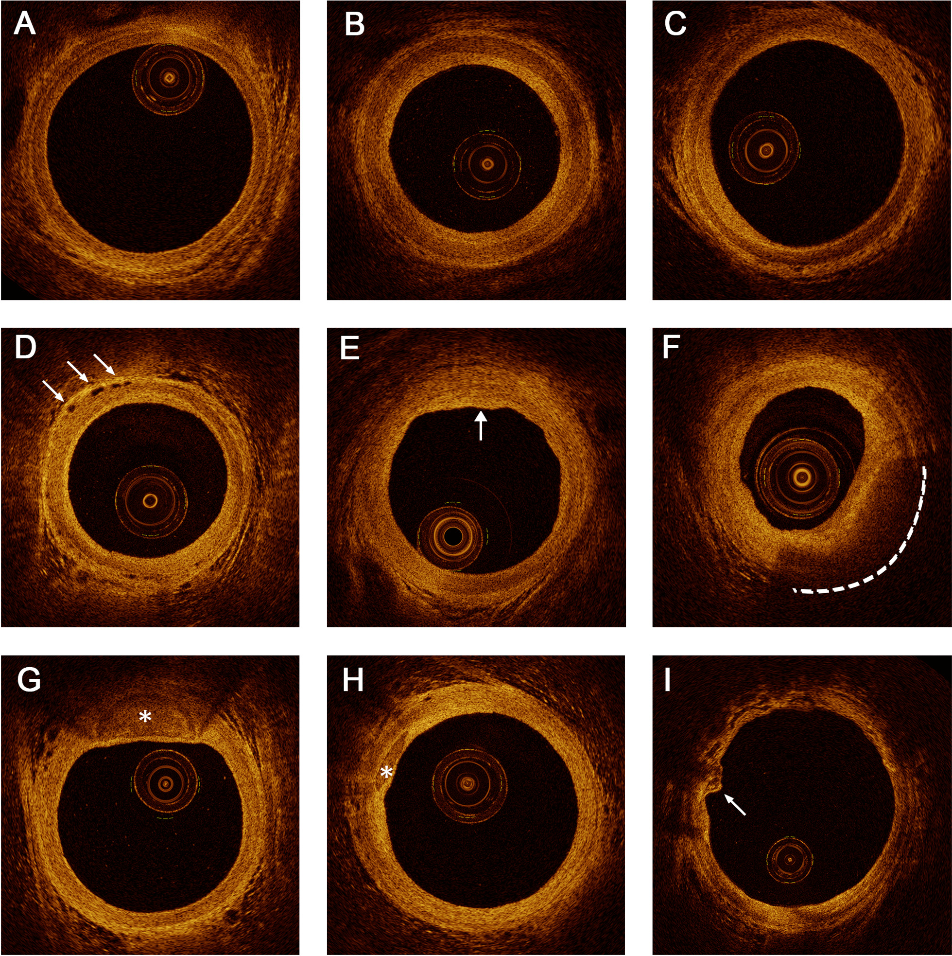
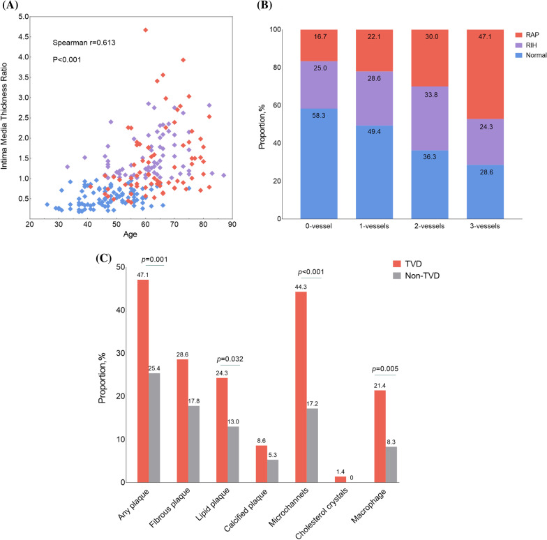
Methods: In this retrospective study involving 239 ACS patients underwent RA OCT without guidewire shadow, 3 groups were divided according to the following criteria: radial artery plaque (RAP) group included patients with fibrous, lipid or calcified plaque; patients without RAP were further classified into radial intimal hyperplasia (RIH) group (intima media thickness ratio [IMR] ≥ 1) or normal group (IMR < 1). The presence and characteristics of RAP and its related risk factors were identified.
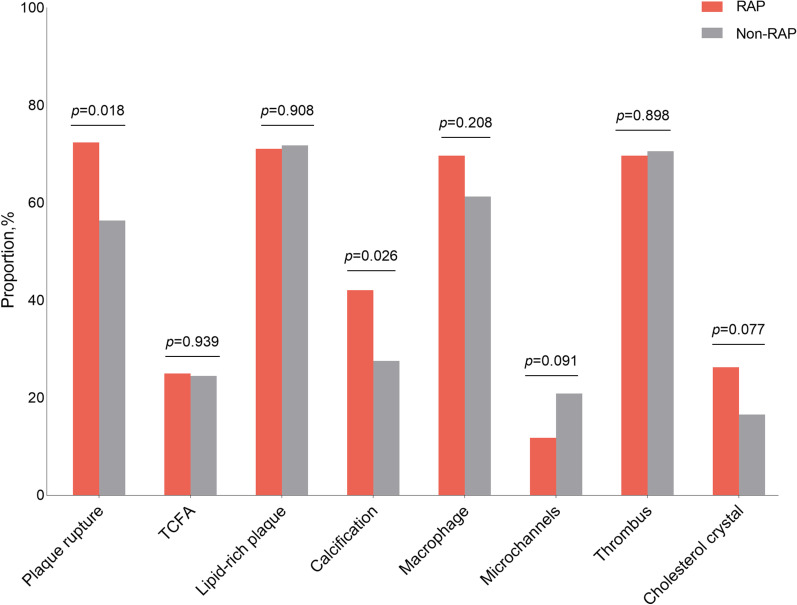
Results: The RAP, RIH and normal groups included 76 (31.8%), 69 (28.9%) and 94 (39.3%) patients, respectively. Patients in RAP group were the oldest, compared with those in the RIH and normal groups (p < 0.001), and more frequently had triple vessel disease (p = 0.004). The percentage of plaque rupture (72.4% vs. 56.4%, p = 0.018) and calcification (42.1% vs. 27.6%, p = 0.026) at culprit lesion were significantly higher in patients with RAP than those without RAP. A total of 148 RAP were revealed by OCT, including fibrous (72, 48.6%), lipid (50, 33.8%) and calcified plaques (26, 17.6%). The microvessels were also frequently observed in the RAP group than that in RIH and normal groups (59.2% vs. 8.7% vs. 9.6%, p < 0.001). Multivariate logistic regression analysis showed that age, diabetes, and smoking history (all p < 0.05) were independent risk factors for RAP.
Conclusions: In terms of insights gained from OCT, RA atherosclerosis is not uncommon in ACS patients by OCT, sharing several morphological characters with early coronary atherosclerosis. Aging, diabetes, and smoking are risk factors for RAP.
Keywords: Atherosclerosis; Optical coherence tomography; Peripheral artery disease; Radial artery.
© 2022. The Author(s).
Conflict of interest statement
All authors declare that they have no competing interests.
Figures
Similar articles
-
Mapping the distribution of radial artery atherosclerosis by optical coherence tomography.BMC Med Imaging. 2025 Feb 13;25(1):47. doi: 10.1186/s12880-025-01583-7. BMC Med Imaging. 2025. PMID: 39948453 Free PMC article.
-
Impact of prediabetes and duration of diabetes on radial artery atherosclerosis in acute coronary syndrome patients: An optical coherence tomography study.Diab Vasc Dis Res. 2022 Jan-Feb;19(1):14791641221078108. doi: 10.1177/14791641221078108. Diab Vasc Dis Res. 2022. PMID: 35184608 Free PMC article.
-
Radial artery intima-media ratio predicts presence of coronary thin-cap fibroatheroma: a frequency domain-optical coherence tomography study.Int J Cardiol. 2013 Oct 3;168(3):1917-22. doi: 10.1016/j.ijcard.2012.12.082. Epub 2013 Jan 29. Int J Cardiol. 2013. PMID: 23369675
-
Utility of optical coherence tomography in acute coronary syndromes.Catheter Cardiovasc Interv. 2023 Jul;102(1):46-55. doi: 10.1002/ccd.30656. Epub 2023 May 28. Catheter Cardiovasc Interv. 2023. PMID: 37245076 Review.
-
What have we learned about plaque rupture in acute coronary syndromes?Curr Cardiol Rep. 2010 Jul;12(4):338-43. doi: 10.1007/s11886-010-0113-x. Curr Cardiol Rep. 2010. PMID: 20425160 Review.
Cited by
-
Radial artery lumen diameter and intima thickness in patients with abdominal aortic aneurysm.JVS Vasc Sci. 2022 Aug 6;3:274-284. doi: 10.1016/j.jvssci.2022.06.001. eCollection 2022. JVS Vasc Sci. 2022. PMID: 36052216 Free PMC article.
-
Mapping the distribution of radial artery atherosclerosis by optical coherence tomography.BMC Med Imaging. 2025 Feb 13;25(1):47. doi: 10.1186/s12880-025-01583-7. BMC Med Imaging. 2025. PMID: 39948453 Free PMC article.
-
Relationship between thrombolysis in myocardial infarction blood flow before percutaneous coronary intervention and the morphological characteristics of culprit vessel plaques in patients with STEMI.Exp Ther Med. 2023 Oct 18;26(6):561. doi: 10.3892/etm.2023.12260. eCollection 2023 Dec. Exp Ther Med. 2023. PMID: 37954121 Free PMC article.
References
-
- Libby P, Buring JE, Badimon L, Hansson GK, Deanfield J, Bittencourt MS, Tokgozoglu L, Lewis EF. Atherosclerosis. Nat Rev Dis Primers. 2019;5(1):56. - PubMed
-
- Eklund C, Omerovic E, Haraldsson I, Friberg P, Gan LM. Radial artery intima-media thickness predicts major cardiovascular events in patients with suspected coronary artery disease. Eur Heart J Cardiovasc Imaging. 2014;15(7):769–775. - PubMed
-
- Xu M, Zhang M, Xu J, Zhu M, Zhang C, Zhang P, Zhang Y. The independent and add-on values of radial intima thickness measured by ultrasound biomicroscopy for diagnosis of coronary artery disease. Eur Heart J Cardiovasc Imaging. 2019;20(8):889–896. - PubMed
-
- Myredal A, Osika W, Li Ming G, Friberg P, Johansson M. Increased intima thickness of the radial artery in patients with coronary heart disease. Vasc Med. 2010;15(1):33–37. - PubMed
-
- Eklund C, Friberg P, Gan LM. High-resolution radial artery intima-media thickness and cardiovascular risk factors in patients with suspected coronary artery disease—comparison with common carotid artery intima-media thickness. Atherosclerosis. 2012;221(1):118–123. - PubMed
Publication types
MeSH terms
Substances
LinkOut - more resources
Full Text Sources
Medical
