. 2022 Mar 21;19(1):69.
doi: 10.1186/s12974-022-02425-x.

The innate immune receptor Nlrp12 suppresses autoimmunity to the retina

Affiliations

The innate immune receptor Nlrp12 suppresses autoimmunity to the retina

Ellen J Lee et al. J Neuroinflammation. .

Abstract

Background: Nod-like receptors (NLRs) are critical to innate immune activation and induction of adaptive T cell responses. Yet, their role in autoinflammatory diseases of the central nervous system (CNS) remains incompletely defined. The NLR, Nlrp12, has been reported to both inhibit and promote neuroinflammation in an animal model of multiple sclerosis (experimental autoimmune encephalomyelitis, EAE), where its T cell-specific role has been investigated. Uveitis resulting from autoimmunity of the neuroretina, an extension of the CNS, involves a breach in immune privilege and entry of T cells into the eye. Here, we examined the contribution of Nlrp12 in a T cell-mediated model of uveitis, experimental autoimmune uveitis (EAU).

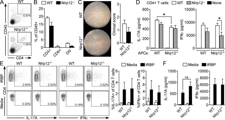
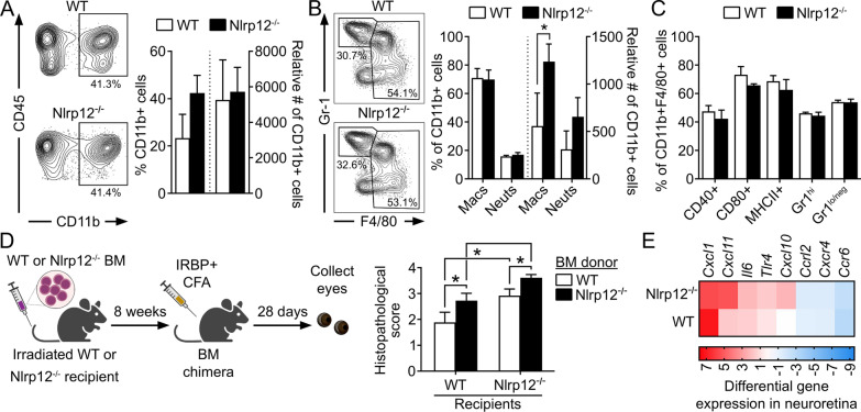
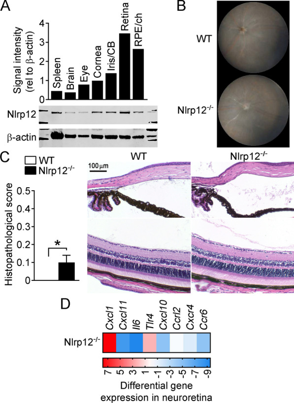
Methods: Mice were immunized with interphotoreceptor retinoid-binding protein peptide 1-20 (IRBP1-20) emulsified in Complete Freund's adjuvant, CFA. Uveitis was evaluated by clinical and histopathological scoring, and comparisons were made in WT vs. Nlrp12-/- mice, lymphopenic Rag1-/- mice reconstituted with WT vs. Nlrp12-/- CD4+ T cells, or among bone marrow (BM) chimeric mice. Antigen-specific Th-effector responses were evaluated by ELISA and intracellular cytokine staining. Cellular composition of uveitic eyes from WT or Nlrp12-/- mice was compared using flow cytometry. Expression of Nlrp12 and of cytokines/chemokines within the neuroretina was evaluated by immunoblotting and quantitative PCR.

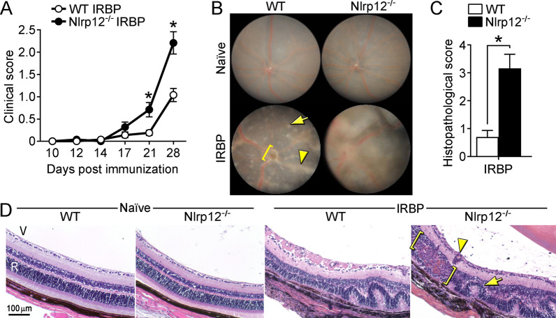
Results: Nlrp12-/- mice developed exacerbated uveitis characterized by extensive vasculitis, chorioretinal infiltrates and photoreceptor damage. Nlrp12 was dispensable for T cell priming and differentiation of peripheral Th1 or Th17 cells, and uveitis in immunodeficient mice reconstituted with either Nlrp12-/- or WT T cells was similar. Collectively, this ruled out T cells as the source of Nlrp12-mediated protection to EAU. Uveitic Nlrp12-/- eyes had more pronounced myeloid cell accumulation than uveitic WT eyes. Transplantation of Nlrp12-/- BM resulted in increased susceptibility to EAU regardless of host genotype, but interestingly, a non-hematopoietic origin for Nlrp12 function was also observed. Indeed, Nlrp12 was found to be constitutively expressed in the neuroretina, where it suppressed chemokine/cytokine induction.

Conclusions: Our data identify a combinatorial role for Nlrp12 in dampening autoimmunity of the neuroretina. These findings could provide a pathway for development of therapies for uveitis and potentially other autoinflammatory/autoimmune diseases of the CNS.

Keywords: Nlrp12; Nod-like receptor; Pattern recognition receptor; Uveitis.


Conflict of interest statement

The authors declare that they have no competing interests.

Figures

Fig. 1
Fig. 1
Nlrp12-deficiency exacerbates autoimmune uveitis: WT and Nlrp12−/− mice were immunized with IRBP1–20 peptide and evaluated for uveitis. A Clinical scoring of eyes (n = 12–13 mice/group combined from 2 studies). B Fundoscopic images of the posterior pole of naïve or at 28 day post-immunization (WT-IRBP = grade 1; Nlrp12−/−-IRBP = grade 3) as compared to naive mice (WT and Nlrp12−/− = grade 0). Corresponding adjuvant-immunized eyes are shown in Fig. 4B. Examples of pathological features include: vasculitis (arrowhead), retinal lesion (arrow), optic nerve head inflammation (bracket). C Histopathology scores of eyes 28 day post-immunization. D Representative photographs of H&E-stained sections of the posterior segment of the eye (approximately 30–60 degrees from the optic nerve head). Examples of pathological features include: retinal fold (arrow), granulomatous formation (bracket), vasculitis (arrowhead). V, vitreous; R, retina. For A and C, data are mean ± SEM, *p < 0.05 by Mann–Whitney
Fig. 2
Fig. 2
Nlrp12-mediated protection against uveitis occurs independent of an inherent T cellular mechanism. The T cell infiltrate in uveitic eyes of WT and Nlrp12−/− mice was evaluated by flow cytometry 21 day post-immunization (A). CD4+ cells were identified from gated live, singles that were CD45+ and identified as CD4+ CD8. Data are of pooled eyes (5 mice/group) and representative of 3 independent experiments. B, C Rag1−/− mice injected with CD4+ T cells purified from naïve, WT or Nlrp12−/− donors. The reconstituted Rag1−/− mice were then immunized for EAU and evaluated for uveitis. Splenocytes of CD4+ T cell-reconstituted Rag1−/− recipients harvested 28 day post-immunization were analyzed by flow cytometry for proportions of indicated T cell subsets (DN, double negative) (B). Clinical uveitis 28 day post-immunization was assessed by fundoscopy. Representative fundus photographs (C, left) and clinical uveitis scores are shown (C, right). D Autologous criss-cross cultures were performed to evaluate Nlrp12 function within APCs in induction of Th1/Th17 immunity. IRBP-reactive CD4+ T cells (purified from immunized donors) were cultured with naïve, WT or Nlrp12−/− APCs, and stimulated with IRBP1–20 peptide. As a negative control, APCs were stimulated with IRBP1–20 peptide in the absence of CD4+ T cells. Cytokine production was measured 18 h later by ELISA. Data are combined from 3 experiments and are mean ± SEM; *p < 0.05. E Th-effector responses were evaluated in IRBP1–20 peptide-stimulated splenocyte cultures from WT vs. Nlrp12−/− mice at 21 d post immunization. Representative dot plot (left) and summary statistics (right, of 3 independent experiments, each with n = 5 mice pooled/group) showing frequencies of IL-17A and IFNγ-producing cells of live CD4+ T cells. F Splenocytes of immunized WT vs. Nlrp12−/− mice were stimulated with IRBP1–20 peptide and cytokine levels in culture supernatants were measured 18 h later by ELISA. Data are mean ± SEM combined from 3 experiments; in each experiment splenocytes from immunized mice were pooled (5 mice/group) and cytokine measured in triplicate wells
Fig. 3
Fig. 3
Nlrp12 suppression of uveitis involves regulation of BM-derived myeloid cellular responses. AC Leukocytic infiltrate in the eyes of WT and Nlrp12−/− mice 21 d post-immunization was evaluated by flow cytometry. Analysis was performed on live singlets expressing the pan-leukocyte marker CD45. A Representative contour plot and summary statistics depicting proportion and number of CD11b+ cells of gated CD45+ cells. B Contour plot and summary statistics of gated CD11b+ cells further distinguished as monocyte–macrophages (being CD45high, CD11clo, F4/80high) vs. neutrophils (CD45high, CD11clo, F4/80loGR-1high). C Expression of activation markers within the gated monocyte/macrophage population. AC Data are mean ± SEM of pooled eyes (5 mice/group) and combined across 3 independent experiments; *p < 0.05 by one-tailed Mann–Whitney. D Bone-marrow (BM)-chimeric mice were generated by transplantation of WT or Nlrp12−/− BM into irradiated WT or Nlrp12−/− recipients. Chimeras were immunized 8 weeks later, and eyes were evaluated for uveitis by histopathology 28 day post-immunization. Data are mean ± SEM (n = 9–10 mice/group combined from 2 experiments), *p < 0.05 by one-tailed Mann–Whitney. E Expression levels of cytokine/chemokine genes in neuroretinas of IRBP1–20-immunized WT and Nlrp12−/− mice (14 day post-immunization, i.e., prior to clinical onset of uveitis) were evaluated by multiplex RT-qPCR. Colors in heatmap indicate the expression level of transcripts relative to control adjuvant-injected WT mice (n = 5 retinas pooled/experimental group)
Fig. 4
Fig. 4
Contribution for Nlrp12 in immune tolerance within the eye. A Nlrp12 expression within distinct ocular tissues of naïve C57BL/6 J mice was assessed by Western blotting. Quantification by densitometry, as shown relative to β-actin expression (top), with representative immunoblot (bottom). B, C Ocular evaluation of adjuvant-injected WT and Nlrp12−/− mice (from experiments presented in Fig. 1 above, n = 8 mice/group). B Representative fundoscopic images and C histopathology scores with representative images, *p < 0.05 by Mann–Whitney. D Expression levels of cytokine/chemokine genes were evaluated by multiplex RT-qPCR in neuroretinas dissected from adjuvant-immunized Nlrp12−/− mice (14 day post-immunization). Colors in heatmap indicate the expression pattern of transcripts relative to adjuvant-injected WT (n = 5 retinas pooled/experimental group)

Similar articles

Cited by

References

    1. Chang JH, Wakefield D. Uveitis: a global perspective. Ocul Immunol Inflamm. 2002;10(4):263–279. - PubMed
    1. Gonzalez MM, Solano MM, Porco TC, Oldenburg CE, Acharya NR, Lin SC, et al. Epidemiology of uveitis in a US population-based study. J Ophthalmic Inflamm Infect. 2018;8(1):6. - PMC - PubMed
    1. Taylor AW, Kaplan HJ. Ocular immune privilege in the year 2010: ocular immune privilege and uveitis. Ocul Immunol Inflamm. 2010;18(6):488–492. - PMC - PubMed
    1. Gery I, Caspi RR. Tolerance induction in relation to the eye. Front Immunol. 2018;9:2304. - PMC - PubMed
    1. Suttorp-Schulten MS, Rothova A. The possible impact of uveitis in blindness: a literature survey. Br J Ophthalmol. 1996;80(9):844–848. - PMC - PubMed

Substances